2ILU

Crystal structure of lactaldehyde dehydrogenase from E. coli: the binary complex with NADPH


Experimental Data Snapshot

  • Method: X-RAY DIFFRACTION
  • Resolution: 2.70 Å
  • R-Value Free: 0.253 
  • R-Value Work: 0.212 

wwPDB Validation   3D Report Full Report


Ligand Structure Quality Assessment 


This is version 1.2 of the entry. See complete history


Literature

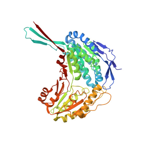
Crystal structure of lactaldehyde dehydrogenase from Escherichia coli and inferences regarding substrate and cofactor specificity.

Di Costanzo, L.Gomez, G.A.Christianson, D.W.

(2007) J Mol Biol 366: 481-493

  • DOI: https://doi.org/10.1016/j.jmb.2006.11.023
  • Primary Citation of Related Structures:  
    2HG2, 2ILU, 2IMP

  • PubMed Abstract: 
  • Aldehyde dehydrogenases catalyze the oxidation of aldehyde substrates to the corresponding carboxylic acids. Lactaldehyde dehydrogenase from Escherichia coli (aldA gene product, P25553) is an NAD(+)-dependent enzyme implicated in the metabolism of l-fucose and l-rhamnose ...

    Aldehyde dehydrogenases catalyze the oxidation of aldehyde substrates to the corresponding carboxylic acids. Lactaldehyde dehydrogenase from Escherichia coli (aldA gene product, P25553) is an NAD(+)-dependent enzyme implicated in the metabolism of l-fucose and l-rhamnose. During the heterologous expression and purification of taxadiene synthase from the Pacific yew, lactaldehyde dehydrogenase from E. coli was identified as a minor (


    Organizational Affiliation

    Roy and Diana Vagelos Laboratories, Department of Chemistry, University of Pennsylvania, Philadelphia, PA 19104-6323, USA.


Macromolecules
Find similar proteins by:  (by identity cutoff)  |  3D Structure
Entity ID: 1
MoleculeChainsSequence LengthOrganismDetailsImage
Aldehyde dehydrogenase AA479Escherichia coliMutation(s): 1 
Gene Names: aldA
EC: 1.2.1.22 (PDB Primary Data), 1.2.1.21 (PDB Primary Data)
UniProt
Find proteins for P25553 (Escherichia coli (strain K12))
Explore P25553 
Go to UniProtKB:  P25553
Entity Groups  
Sequence Clusters30% Identity50% Identity70% Identity90% Identity95% Identity100% Identity
UniProt GroupP25553
Protein Feature View
Expand
  • Reference Sequence
Small Molecules
Ligands 2 Unique
IDChainsName / Formula / InChI Key2D Diagram3D Interactions
NDP
Query on NDP

Download Ideal Coordinates CCD File 
D [auth A]NADPH DIHYDRO-NICOTINAMIDE-ADENINE-DINUCLEOTIDE PHOSPHATE
C21 H30 N7 O17 P3
ACFIXJIJDZMPPO-NNYOXOHSSA-N
 Ligand Interaction
SO4
Query on SO4

Download Ideal Coordinates CCD File 
B [auth A],
C [auth A]
SULFATE ION
O4 S
QAOWNCQODCNURD-UHFFFAOYSA-L
 Ligand Interaction
Modified Residues  1 Unique
IDChainsTypeFormula2D DiagramParent
OCS
Query on OCS
A L-PEPTIDE LINKINGC3 H7 N O5 SCYS
Experimental Data & Validation

Experimental Data

  • Method: X-RAY DIFFRACTION
  • Resolution: 2.70 Å
  • R-Value Free: 0.253 
  • R-Value Work: 0.212 
  • Space Group: P 64 2 2
Unit Cell:
Length ( Å )Angle ( ˚ )
a = 143.566α = 90
b = 143.566β = 90
c = 108.763γ = 120
Software Package:
Software NamePurpose
HKL-2000data collection
CNSrefinement
HKL-2000data reduction
SCALEPACKdata scaling
CNSphasing

Structure Validation

View Full Validation Report


Ligand Structure Quality Assessment 


Entry History 

Deposition Data

Revision History  (Full details and data files)

  • Version 1.0: 2007-05-08
    Type: Initial release
  • Version 1.1: 2008-05-01
    Changes: Version format compliance
  • Version 1.2: 2011-07-13
    Changes: Derived calculations, Version format compliance