


Crystal Structure of the SAM Dependent Methyltransferase from Agrobacterium tumefaciens
Kim, Y., Joachimiak, A., Xu, X., Gu, J., Edwards, A., Savchenko, A.To be published.
Experimental Data Snapshot
Entity ID: 1 | |||||
|---|---|---|---|---|---|
| Molecule | Chains | Sequence Length | Organism | Details | Image |
| SAM dependent methyltransferase | 332 | Agrobacterium fabrum str. C58 | Mutation(s): 7 | 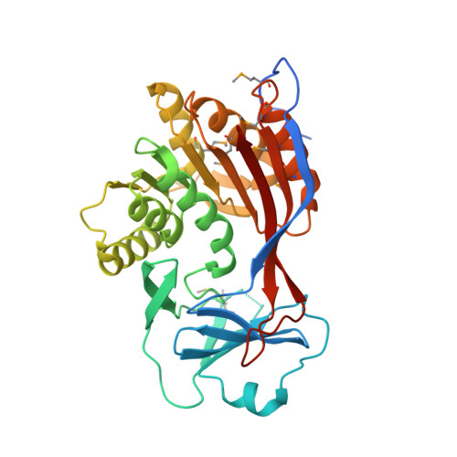 | |
UniProt | |||||
Entity Groups | |||||
| Sequence Clusters | 30% Identity50% Identity70% Identity90% Identity95% Identity100% Identity | ||||
| UniProt Group | A9CKD6 | ||||
Sequence AnnotationsExpand | |||||
Reference Sequence | |||||
| Ligands 4 Unique | |||||
|---|---|---|---|---|---|
| ID | Chains | Name / Formula / InChI Key | 2D Diagram | 3D Interactions | |
| SAM Download:Ideal Coordinates CCD File | D [auth A], J [auth B], N [auth C] | S-ADENOSYLMETHIONINE C15 H22 N6 O5 S MEFKEPWMEQBLKI-FCKMPRQPSA-N |  | ||
| GOL Download:Ideal Coordinates CCD File | E [auth A], O [auth C] | GLYCEROL C3 H8 O3 PEDCQBHIVMGVHV-UHFFFAOYSA-N |  | ||
| ACY Download:Ideal Coordinates CCD File | F [auth A] G [auth A] H [auth A] P [auth C] Q [auth C] | ACETIC ACID C2 H4 O2 QTBSBXVTEAMEQO-UHFFFAOYSA-N |  | ||
| FMT Download:Ideal Coordinates CCD File | I [auth A] K [auth B] L [auth B] M [auth B] S [auth C] | FORMIC ACID C H2 O2 BDAGIHXWWSANSR-UHFFFAOYSA-N |  | ||
| Modified Residues 1 Unique | |||||
|---|---|---|---|---|---|
| ID | Chains | Type | Formula | 2D Diagram | Parent |
| MSE Query on MSE | A, B, C | L-PEPTIDE LINKING | C5 H11 N O2 Se |  | MET |
| Length ( Å ) | Angle ( ˚ ) |
|---|---|
| a = 83.941 | α = 90 |
| b = 145.555 | β = 90 |
| c = 204.895 | γ = 90 |
| Software Name | Purpose |
|---|---|
| REFMAC | refinement |
| SBC-Collect | data collection |
| HKL-2000 | data collection |
| HKL-2000 | data reduction |
| HKL-2000 | data scaling |
| HKL-3000 | phasing |
| SHELXCD | phasing |
| SHELXD | phasing |
| SHELXE | model building |
| SOLVE | phasing |
| RESOLVE | phasing |
| MLPHARE | phasing |
| Coot | model building |
| ARP/wARP | model building |


