Structural determinants in the group III truncated hemoglobin from Campylobacter jejuni.
Nardini, M., Pesce, A., Labarre, M., Richard, C., Bolli, A., Ascenzi, P., Guertin, M., Bolognesi, M.(2006) J Biological Chem 281: 37803-37812
- PubMed: 17023416 Search on PubMed
- DOI: https://doi.org/10.1074/jbc.M607254200
- Primary Citation Related Structures:
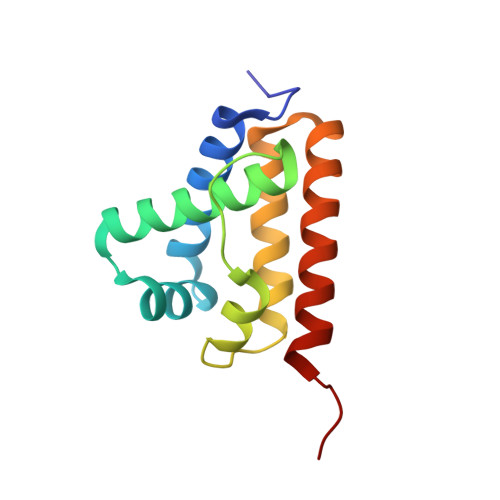
2IG3 - PubMed Abstract:
Truncated hemoglobins (trHbs) constitute a distinct lineage in the globin superfamily, distantly related in size and fold to myoglobin and monomeric hemoglobins. Their phylogenetic analyses revealed that three groups (I, II, and III) compose the trHb family. Group I and II trHbs adopt a simplified globin fold, essentially composed of a 2-on-2 alpha-helical sandwich, wrapped around the heme group. So far no structural data have been reported for group III trHbs. Here we report the three-dimensional structure of the group III trHbP from the eubacterium Campylobacter jejuni. The 2.15-A resolution crystal structure of C. jejuni trHbP (cyano-met form) shows that the 2-on-2 trHb fold is substantially conserved in the trHb group III, despite the absence of the Gly-based sequence motifs that were considered necessary for the attainment of the trHb specific fold. The heme crevice presents important structural modifications in the C-E region and in the FG helical hinge, with novel surface clefts at the proximal heme site. Contrary to what has been observed for group I and II trHbs, no protein matrix tunnel/cavity system is evident in C. jejuni trHbP. A gating movement of His(E7) side chain (found in two alternate conformations in the crystal structure) may be instrumental for ligand entry to the heme distal site. Sequence conservation allows extrapolating part of the structural results here reported to the whole trHb group III.
- Department of Biomolecular Sciences and Biotechnology, and CNR-INFM, University of Milano, I-20131 Milano, Italy.
Organizational Affiliation: