2IF7 | pdb_00002if7

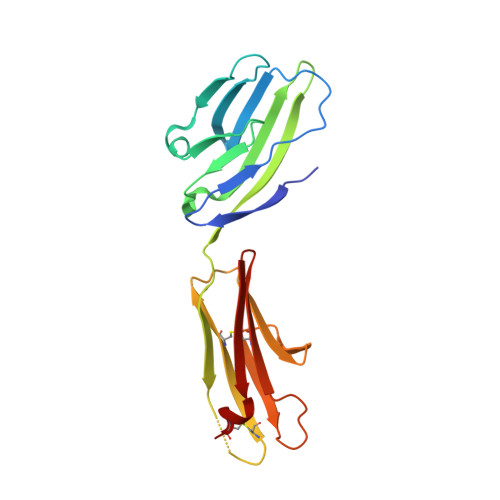
Crystal Structure of NTB-A


Experimental Data Snapshot

  • Method: X-RAY DIFFRACTION
  • Resolution: 3.00 Å
  • R-Value Free: 
    0.266 (Depositor), 0.260 (DCC) 
  • R-Value Work: 
    0.213 (Depositor), 0.208 (DCC) 
  • R-Value Observed: 
    0.216 (Depositor) 

wwPDB Validation 3D Report Full Report

Validation slider image for 2IF7

This is version 1.4 of the entry. See complete history. 

Literature

NTB-A Receptor Crystal Structure: Insights into Homophilic Interactions in the Signaling Lymphocytic Activation Molecule Receptor Family.

Cao, E., Ramagopal, U.A., Fedorov, A., Fedorov, E., Yan, Q., Lary, J.W., Cole, J.L., Nathenson, S.G., Almo, S.C.

(2006) Immunity 25: 559-570

  • DOI: https://doi.org/10.1016/j.immuni.2006.06.020
  • Primary Citation Related Structures: 
    2IF7

  • PubMed Abstract: 

    The signaling lymphocytic activation molecule (SLAM) family includes homophilic and heterophilic receptors that regulate both innate and adaptive immunity. The ectodomains of most SLAM family members are composed of an N-terminal IgV domain and a C-terminal IgC2 domain. NK-T-B-antigen (NTB-A) is a homophilic receptor that stimulates cytotoxicity in natural killer (NK) cells, regulates bactericidal activities in neutrophils, and potentiates T helper 2 (Th2) responses. The 3.0 A crystal structure of the complete NTB-A ectodomain revealed a rod-like monomer that self-associates to form a highly kinked dimer spanning an end-to-end distance of approximately 100 A. The NTB-A homophilic and CD2-CD58 heterophilic dimers show overall structural similarities but differ in detailed organization and physicochemical properties of their respective interfaces. The NTB-A structure suggests a mechanism responsible for binding specificity within the SLAM family and imposes physical constraints relevant to the colocalization of SLAM-family proteins with other signaling molecules in the immunological synapse.


  • Organizational Affiliation: 
    • Department of Cell Biology, Albert Einstein College of Medicine, Bronx, New York 10461, USA.

Macromolecule Content 

  • Total Structure Weight: 86.86 kDa 
  • Atom Count: 5,966 
  • Modeled Residue Count: 757 
  • Deposited Residue Count: 772 
  • Unique protein chains: 1

Macromolecules

Find similar proteins by:|  3D Structure
Entity ID: 1
MoleculeChains  Sequence LengthOrganismDetailsImage
SLAM family member 6
A, B, C, D
193Homo sapiensMutation(s): 0 
Gene Names: SLAMF6, KALI
UniProt & NIH Common Fund Data Resources
Find proteins for Q96DU3 (Homo sapiens)
Explore Q96DU3 
Go to UniProtKB:  Q96DU3
PHAROS:  Q96DU3
GTEx:  ENSG00000162739 
Entity Groups
Sequence Clusters30% Identity50% Identity70% Identity90% Identity95% Identity100% Identity
UniProt GroupQ96DU3
Sequence Annotations
Expand
Reference Sequence

Experimental Data & Validation

Experimental Data

  • Method: X-RAY DIFFRACTION
  • Resolution: 3.00 Å
  • R-Value Free:  0.266 (Depositor), 0.260 (DCC) 
  • R-Value Work:  0.213 (Depositor), 0.208 (DCC) 
  • R-Value Observed: 0.216 (Depositor) 
Space Group: C 1 2 1
Unit Cell:
Length ( Å )Angle ( ˚ )
a = 121.196α = 90
b = 148.452β = 112.86
c = 86.572γ = 90
Software Package:
Software NamePurpose
DENZOdata reduction
SCALEPACKdata scaling
REFMACrefinement
PDB_EXTRACTdata extraction
CBASSdata collection
HKL-2000data reduction
SHELXDphasing
SHELXEmodel building
SOLVEphasing
RESOLVEphasing
Cootmodel building
Omodel building

Structure Validation

View Full Validation Report


Entry History 

Deposition Data

Revision History  (Full details and data files)

  • Version 1.0: 2006-10-17
    Type: Initial release
  • Version 1.1: 2008-05-01
    Changes: Version format compliance
  • Version 1.2: 2011-07-13
    Changes: Advisory, Version format compliance
  • Version 1.3: 2017-10-18
    Changes: Refinement description
  • Version 1.4: 2024-11-06
    Changes: Data collection, Database references, Derived calculations, Structure summary