2HZY | pdb_00002hzy

Mouse fumarylacetoacetate hydrolase complexes with a transition-state mimic of the complete substrate


Experimental Data Snapshot

  • Method: X-RAY DIFFRACTION
  • Resolution: 1.35 Å
  • R-Value Free: 
    0.188 (Depositor), 0.189 (DCC) 
  • R-Value Work: 
    0.168 (Depositor), 0.169 (DCC) 
  • R-Value Observed: 
    0.169 (Depositor) 

Starting Model: experimental
View more details

wwPDB Validation 3D Report Full Report

Validation slider image for 2HZY

Ligand Structure Quality Assessment 


This is version 1.3 of the entry. See complete history

Literature

Slow-onset inhibition of fumarylacetoacetate hydrolase by phosphinate mimics of the tetrahedral intermediate: kinetics, crystal structure and pharmacokinetics.

Bateman, R.L.Ashworth, J.Witte, J.F.Baker, L.J.Bhanumoorthy, P.Timm, D.E.Hurley, T.D.Grompe, M.McClard, R.W.

(2007) Biochem J 402: 251-260

  • DOI: https://doi.org/10.1042/BJ20060961
  • Primary Citation Related Structures: 
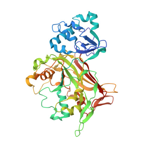
    2HZY

  • PubMed Abstract: 

    FAH (fumarylacetoacetate hydrolase) catalyses the final step of tyrosine catabolism to produce fumarate and acetoacetate. HT1 (hereditary tyrosinaemia type 1) results from deficiency of this enzyme. Previously, we prepared a partial mimic of the putative tetrahedral intermediate in the reaction catalysed by FAH co-crystallized with the enzyme to reveal details of the mechanism [Bateman, Bhanumoorthy, Witte, McClard, Grompe and Timm (2001) J. Biol. Chem. 276, 15284-15291]. We have now successfully synthesized complete mimics CEHPOBA {4-[(2-carboxyethyl)-hydroxyphosphinyl]-3-oxobutyrate} and COPHPAA {3-[(3-carboxy-2-oxopropyl)hydroxyphosphinyl]acrylate}, which inhibit FAH in slow-onset tight-binding mode with K(i) values of 41 and 12 nM respectively. A high-resolution (1.35 A; 1 A=0.1 nm) crystal structure of the FAH.CEHPOBA complex was solved to reveal the affinity determinants for these compounds and to provide further insight into the mechanism of FAH catalysis. These compounds are active in vivo, and CEHPOBA demonstrated a notable dose-dependent increase in SA (succinylacetone; a metabolite seen in patients with HT1) in mouse serum after repeated injections, and, following a single injection (1 mumol/g; intraperitoneal), only a modest regain of FAH enzyme activity was detected in liver protein isolates after 24 h. These potent inhibitors provide a means to chemically phenocopy the metabolic defects of either HT1 or FAH knockout mice and promise future pharmacological utility for hepatocyte transplantation.


  • Organizational Affiliation
    • Arthur F. Scott Laboratory of Chemistry, Reed College, 3203 SE Woodstock Blvd, Portland, OR 97202, USA.

Macromolecule Content 

  • Total Structure Weight: 93.85 kDa 
  • Atom Count: 7,532 
  • Modeled Residue Count: 834 
  • Deposited Residue Count: 842 
  • Unique protein chains: 1

Macromolecules

Find similar proteins by:|  3D Structure
Entity ID: 1
MoleculeChains  Sequence LengthOrganismDetailsImage
Fumarylacetoacetase
A, B
421Mus musculusMutation(s): 0 
Gene Names: Fah
EC: 3.7.1.2
UniProt & NIH Common Fund Data Resources
Find proteins for P35505 (Mus musculus)
Explore P35505 
Go to UniProtKB:  P35505
IMPC:  MGI:95482
Entity Groups
Sequence Clusters30% Identity50% Identity70% Identity90% Identity95% Identity100% Identity
UniProt GroupP35505
Sequence Annotations
Expand
Reference Sequence

Small Molecules

Ligands 5 Unique
IDChains Name / Formula / InChI Key2D Diagram3D Interactions
DHJ

Query on DHJ


Download:Ideal Coordinates CCD File
K [auth A],
S [auth B]
4-(2-CARBOXYETHYL)(HYDROXY)PHOSPHORYL]-3-OXOBUTANOIC ACID
C7 H11 O7 P
YKFFQOHPUCXFLE-UHFFFAOYSA-N
NI

Query on NI


Download:Ideal Coordinates CCD File
F [auth A]
G [auth A]
H [auth A]
I [auth A]
J [auth A]
F [auth A],
G [auth A],
H [auth A],
I [auth A],
J [auth A],
N [auth B],
O [auth B],
P [auth B],
Q [auth B],
R [auth B]
NICKEL (II) ION
Ni
VEQPNABPJHWNSG-UHFFFAOYSA-N
MN

Query on MN


Download:Ideal Coordinates CCD File
D [auth A],
M [auth B]
MANGANESE (II) ION
Mn
WAEMQWOKJMHJLA-UHFFFAOYSA-N
CA

Query on CA


Download:Ideal Coordinates CCD File
E [auth A]CALCIUM ION
Ca
BHPQYMZQTOCNFJ-UHFFFAOYSA-N
NA

Query on NA


Download:Ideal Coordinates CCD File
C [auth A],
L [auth B]
SODIUM ION
Na
FKNQFGJONOIPTF-UHFFFAOYSA-N

Experimental Data & Validation

Experimental Data

  • Method: X-RAY DIFFRACTION
  • Resolution: 1.35 Å
  • R-Value Free:  0.188 (Depositor), 0.189 (DCC) 
  • R-Value Work:  0.168 (Depositor), 0.169 (DCC) 
  • R-Value Observed: 0.169 (Depositor) 
Space Group: P 1 21 1
Unit Cell:
Length ( Å )Angle ( ˚ )
a = 64.144α = 90
b = 109.472β = 102.35
c = 67.491γ = 90
Software Package:
Software NamePurpose
REFMACrefinement
HKL-2000data collection
HKL-2000data reduction
SCALEPACKdata scaling
AMoREphasing

Structure Validation

View Full Validation Report


Ligand Structure Quality Assessment 


Entry History 

Deposition Data

Revision History  (Full details and data files)

  • Version 1.0: 2006-08-22
    Type: Initial release
  • Version 1.1: 2008-05-01
    Changes: Version format compliance
  • Version 1.2: 2011-07-13
    Changes: Version format compliance
  • Version 1.3: 2023-08-30
    Changes: Data collection, Database references, Derived calculations, Refinement description