Structure and stability of designed TPR protein superhelices: unusual crystal packing and implications for natural TPR proteins.
Kajander, T., Cortajarena, A.L., Mochrie, S., Regan, L.(2007) Acta Crystallogr D Biol Crystallogr 63: 800-811
- PubMed: 17582171 Search on PubMed
- DOI: https://doi.org/10.1107/S0907444907024353
- Primary Citation Related Structures:
2AVP, 2FO7, 2HYZ - PubMed Abstract:
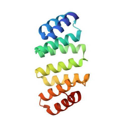
The structure and stability of repeat proteins has been little studied in comparison to the properties of the more familiar globular proteins. Here, the structure and stability of designed tetratricopeptide-repeat (TPR) proteins is described. The TPR is a 34-amino-acid motif which adopts a helix-turn-helix structure and occurs as tandem repeats. The design of a consensus TPR motif (CTPR) has previously been described. Here, the crystal structures and stabilities of proteins that contain eight or 20 identical tandem repeats of the CTPR motif (CTPR8 and CTPR20) are presented. Both CTPR8 and CTPR20 adopt a superhelical overall structure. The structures of the different-length CTPR proteins are compared with each other and with the structures of natural TPR domains. Also, the unusual and perhaps unique crystal-packing interactions resulting in pseudo-infinite crystalline superhelices observed in the different crystal forms of CTPR8 and CTPR20 are discussed. Finally, it is shown that the thermodynamic behavior of CTPR8 and CTPR20 can be predicted from the behavior of other TPRs in this series using an Ising model-based analysis. The designed protein series CTPR2-CTPR20 covers the natural size repertoire of TPR domains and as such is an excellent model system for natural TPR proteins.
- Department of Molecular Biophysics and Biochemistry, Yale University, New Haven, USA.
Organizational Affiliation: