2HTH | pdb_00002hth

Structural basis for ubiquitin recognition by the human EAP45/ESCRT-II GLUE domain


Experimental Data Snapshot

  • Method: X-RAY DIFFRACTION
  • Resolution: 2.70 Å
  • R-Value Free: 
    0.289 (Depositor), 0.292 (DCC) 
  • R-Value Work: 
    0.248 (Depositor), 0.244 (DCC) 
  • R-Value Observed: 
    0.252 (Depositor) 

wwPDB Validation 3D Report Full Report

Validation slider image for 2HTH

This is version 1.4 of the entry. See complete history

Literature

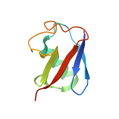
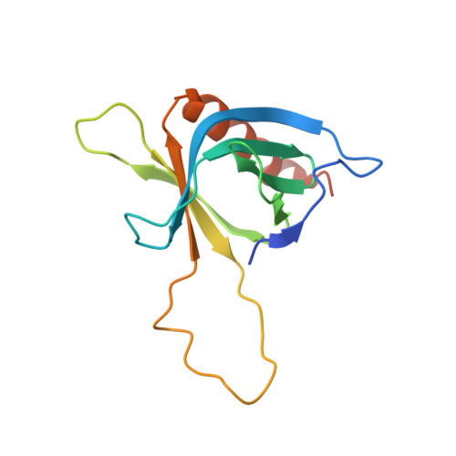
Structural basis for ubiquitin recognition by the human ESCRT-II EAP45 GLUE domain.

Alam, S.L.Langelier, C.Whitby, F.G.Koirala, S.Robinson, H.Hill, C.P.Sundquist, W.I.

(2006) Nat Struct Mol Biol 13: 1029-1030

  • DOI: https://doi.org/10.1038/nsmb1160
  • Primary Citation Related Structures: 
    2HTH

  • PubMed Abstract: 

    The ESCRT-I and ESCRT-II complexes help sort ubiquitinated proteins into vesicles that accumulate within multivesicular bodies (MVBs). Crystallographic and biochemical analyses reveal that the GLUE domain of the human ESCRT-II EAP45 (also called VPS36) subunit is a split pleckstrin-homology domain that binds ubiquitin along one edge of the beta-sandwich. The structure suggests how human ESCRT-II can couple recognition of ubiquitinated cargoes and endosomal phospholipids during MVB protein sorting.


  • Organizational Affiliation
    • Department of Biochemistry, University of Utah, Salt Lake City, Utah 84112-5650, USA.

Macromolecule Content 

  • Total Structure Weight: 24.83 kDa 
  • Atom Count: 1,640 
  • Modeled Residue Count: 202 
  • Deposited Residue Count: 216 
  • Unique protein chains: 2

Macromolecules

Find similar proteins by:|  3D Structure
Entity ID: 1
MoleculeChains  Sequence LengthOrganismDetailsImage
Ubiquitin76Homo sapiensMutation(s): 0 
Gene Names: RPS27AUBA52UBBUBC
UniProt & NIH Common Fund Data Resources
Find proteins for P0CG48 (Homo sapiens)
Explore P0CG48 
Go to UniProtKB:  P0CG48
PHAROS:  P0CG48
GTEx:  ENSG00000150991 
Entity Groups
Sequence Clusters30% Identity50% Identity70% Identity90% Identity95% Identity100% Identity
UniProt GroupP0CG48
Sequence Annotations
Expand
Reference Sequence
Find similar proteins by:|  3D Structure
Entity ID: 2
MoleculeChains  Sequence LengthOrganismDetailsImage
Vacuolar protein sorting protein 36140Homo sapiensMutation(s): 0 
Gene Names: VPS36C13orf9EAP45
UniProt & NIH Common Fund Data Resources
Find proteins for Q86VN1 (Homo sapiens)
Explore Q86VN1 
Go to UniProtKB:  Q86VN1
PHAROS:  Q86VN1
GTEx:  ENSG00000136100 
Entity Groups
Sequence Clusters30% Identity50% Identity70% Identity90% Identity95% Identity100% Identity
UniProt GroupQ86VN1
Sequence Annotations
Expand
Reference Sequence

Experimental Data & Validation

Experimental Data

  • Method: X-RAY DIFFRACTION
  • Resolution: 2.70 Å
  • R-Value Free:  0.289 (Depositor), 0.292 (DCC) 
  • R-Value Work:  0.248 (Depositor), 0.244 (DCC) 
  • R-Value Observed: 0.252 (Depositor) 
Space Group: P 42 21 2
Unit Cell:
Length ( Å )Angle ( ˚ )
a = 102.727α = 90
b = 102.727β = 90
c = 54.306γ = 90
Software Package:
Software NamePurpose
DENZOdata reduction
SCALEPACKdata scaling
SOLVEphasing
RESOLVEphasing
REFMACrefinement
PDB_EXTRACTdata extraction

Structure Validation

View Full Validation Report


Entry History 

Deposition Data

Revision History  (Full details and data files)

  • Version 1.0: 2006-10-24
    Type: Initial release
  • Version 1.1: 2008-05-01
    Changes: Version format compliance
  • Version 1.2: 2011-07-13
    Changes: Version format compliance
  • Version 1.3: 2017-10-18
    Changes: Advisory, Refinement description
  • Version 1.4: 2024-02-14
    Changes: Advisory, Data collection, Database references