2HT4 | pdb_00002ht4

Structure of the Escherichia coli ClC chloride channel Y445W mutant and Fab complex


Experimental Data Snapshot

  • Method: X-RAY DIFFRACTION
  • Resolution: 3.20 Å
  • R-Value Free: 
    0.261 (Depositor), 0.253 (DCC) 
  • R-Value Work: 
    0.239 (Depositor), 0.231 (DCC) 
  • R-Value Observed: 
    0.240 (Depositor) 

wwPDB Validation 3D Report Full Report

Validation slider image for 2HT4

This is version 1.4 of the entry. See complete history

Literature

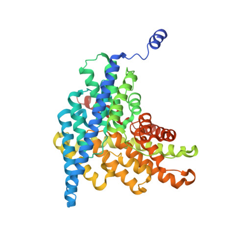
Synergism Between Halide Binding and Proton Transport in a CLC-type Exchanger.

Accardi, A.Lobet, S.Williams, C.Miller, C.Dutzler, R.

(2006) J Mol Biology 362: 691-699

  • DOI: https://doi.org/10.1016/j.jmb.2006.07.081
  • Primary Citation Related Structures: 
    2HLF, 2HT2, 2HT3, 2HT4, 2HTK, 2HTL

  • PubMed Abstract: 

    The Cl-/H+ exchange-transporter CLC-ec1 mediates stoichiometric transmembrane exchange of two Cl- ions for one proton. A conserved tyrosine residue, Y445, coordinates one of the bound Cl- ions visible in the structure of this protein and is located near the intersection of the Cl- and H+ pathways. Mutants of this tyrosine were scrutinized for effects on the coupled transport of Cl- and H+ determined electrophysiologically and on protein structure determined crystallographically. Despite the strong conservation of Y445 in the CLC family, substitution of F or W at this position preserves wild-type transport behavior. Substitution by A, E, or H, however, produces uncoupled proteins with robust Cl- transport but greatly impaired movement of H+. The obligatory 2 Cl-/1 H+ stoichiometry is thus lost in these mutants. The structures of all the mutants are essentially identical to wild-type, but apparent anion occupancy in the Cl- binding region correlates with functional H+ coupling. In particular, as determined by anomalous diffraction in crystals grown in Br-, an electrophysiologically competent Cl- analogue, the well-coupled transporters show strong Br- electron density at the "inner" and "central" Cl- binding sites. However, in the uncoupled mutants, Br- density is absent at the central site, while still present at the inner site. An additional mutant, Y445L, is intermediate in both functional and structural features. This mutant clearly exchanges H+ for Cl-, but at a reduced H+-to-Cl- ratio; likewise, both the central and inner sites are occupied by Br-, but the central site shows lower Br- density than in wild-type (or in Y445F,W). The correlation between proton coupling and central-site occupancy argues that halide binding to the central transport site somehow facilitates movement of H+, a synergism that is not readily understood in terms of alternating-site antiport schemes.


  • Organizational Affiliation
    • Department of Biochemistry, Howard Hughes Medical Institute, Brandeis University, Waltham, MA 02454-9110, USA.

Macromolecule Content 

  • Total Structure Weight: 194.71 kDa 
  • Atom Count: 13,231 
  • Modeled Residue Count: 1,749 
  • Deposited Residue Count: 1,810 
  • Unique protein chains: 3

Macromolecules

Find similar proteins by:|  3D Structure
Entity ID: 1
MoleculeChains  Sequence LengthOrganismDetailsImage
H(+)/Cl(-) exchange transporter clcA
A, B
473Escherichia coliMutation(s): 1 
Gene Names: clcAeriC
Membrane Entity: Yes 
UniProt
Find proteins for P37019 (Escherichia coli (strain K12))
Explore P37019 
Go to UniProtKB:  P37019
Entity Groups
Sequence Clusters30% Identity50% Identity70% Identity90% Identity95% Identity100% Identity
UniProt GroupP37019
Sequence Annotations
Expand
Reference Sequence
Find similar proteins by:|  3D Structure
Entity ID: 2
MoleculeChains  Sequence LengthOrganismDetailsImage
Fab fragment, Heavy chain
C, E
221Mus musculusMutation(s): 0 
UniProt
Find proteins for P01808 (Mus musculus)
Explore P01808 
Go to UniProtKB:  P01808
Entity Groups
Sequence Clusters30% Identity50% Identity70% Identity90% Identity95% Identity100% Identity
UniProt GroupP01808
Sequence Annotations
Expand
Reference Sequence
Find similar proteins by:|  3D Structure
Entity ID: 3
MoleculeChains  Sequence LengthOrganismDetailsImage
Fab fragment, Light chain
D, F
211Mus musculusMutation(s): 0 

Experimental Data & Validation

Experimental Data

  • Method: X-RAY DIFFRACTION
  • Resolution: 3.20 Å
  • R-Value Free:  0.261 (Depositor), 0.253 (DCC) 
  • R-Value Work:  0.239 (Depositor), 0.231 (DCC) 
  • R-Value Observed: 0.240 (Depositor) 
Space Group: C 1 2 1
Unit Cell:
Length ( Å )Angle ( ˚ )
a = 234.007α = 90
b = 95.94β = 133.2
c = 175.327γ = 90
Software Package:
Software NamePurpose
REFMACrefinement
DENZOdata reduction
SCALEPACKdata scaling
CNSphasing

Structure Validation

View Full Validation Report


Entry History 

Deposition Data

Revision History  (Full details and data files)

  • Version 1.0: 2006-09-19
    Type: Initial release
  • Version 1.1: 2008-05-01
    Changes: Version format compliance
  • Version 1.2: 2011-07-13
    Changes: Version format compliance
  • Version 1.3: 2021-10-20
    Changes: Database references, Derived calculations
  • Version 1.4: 2024-10-30
    Changes: Data collection, Refinement description, Structure summary