The first three domains of the insulin receptor differ structurally from the insulin-like growth factor 1 receptor in the regions governing ligand specificity.
Lou, M., Garrett, T.P., McKern, N.M., Hoyne, P.A., Epa, V.C., Bentley, J.D., Lovrecz, G.O., Cosgrove, L.J., Frenkel, M.J., Ward, C.W.(2006) Proc Natl Acad Sci U S A 103: 12429-12434
- PubMed: 16894147 Search on PubMedSearch on PubMed Central
- DOI: https://doi.org/10.1073/pnas.0605395103
- Primary Citation Related Structures:
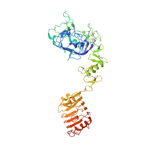
2HR7 - PubMed Abstract:
The insulin receptor (IR) and the type-1 insulin-like growth factor receptor (IGF1R) are homologous multidomain proteins that bind insulin and IGF with differing specificity. Here we report the crystal structure of the first three domains (L1-CR-L2) of human IR at 2.3 A resolution and compare it with the previously determined structure of the corresponding fragment of IGF1R. The most important differences seen between the two receptors are in the two regions governing ligand specificity. The first is at the corner of the ligand-binding surface of the L1 domain, where the side chain of F39 in IR forms part of the ligand binding surface involving the second (central) beta-sheet. This is very different to the location of its counterpart in IGF1R, S35, which is not involved in ligand binding. The second major difference is in the sixth module of the CR domain, where IR contains a larger loop that protrudes further into the ligand-binding pocket. This module, which governs IGF1-binding specificity, shows negligible sequence identity, significantly more alpha-helix, an additional disulfide bond, and opposite electrostatic potential compared to that of the IGF1R.
- Division of Molecular and Health Technologies, Commonwealth Scientific and Industrial Research Organization, 343 Royal Parade, Parkville, Victoria 3052, Australia.
Organizational Affiliation: