


Crystal structure of iron bound Rubrerythrin from Pyrococcus Furiosus
Dillard, B.D., Clarkson, S.M., Strand, K.R., Ruble, J.R., Chen, L., Liu, Z.J., Jenney Jr., F.E., Adams, M.W.W., Rose, J.P., Wang, B.C.To be published.
Experimental Data Snapshot
Starting Model: experimental
View more details
wwPDB Validation 3D Report Full Report

Entity ID: 1 | |||||
|---|---|---|---|---|---|
| Molecule | Chains | Sequence Length | Organism | Details | Image |
| Rubrerythrin | 171 | Pyrococcus furiosus | Mutation(s): 0 | 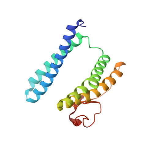 | |
UniProt | |||||
Entity Groups | |||||
| Sequence Clusters | 30% Identity50% Identity70% Identity90% Identity95% Identity100% Identity | ||||
| UniProt Group | Q9UWP7 | ||||
Sequence AnnotationsExpand | |||||
Reference Sequence | |||||
| Ligands 1 Unique | |||||
|---|---|---|---|---|---|
| ID | Chains | Name / Formula / InChI Key | 2D Diagram | 3D Interactions | |
| FE Download:Ideal Coordinates CCD File | C [auth A] D [auth A] E [auth A] F [auth B] G [auth B] | FE (III) ION Fe VTLYFUHAOXGGBS-UHFFFAOYSA-N |  | ||
| Length ( Å ) | Angle ( ˚ ) |
|---|---|
| a = 105.402 | α = 90 |
| b = 105.402 | β = 90 |
| c = 80.095 | γ = 90 |
| Software Name | Purpose |
|---|---|
| CCP4 | model building |
| REFMAC | refinement |
| HKL-2000 | data reduction |
| HKL-2000 | data scaling |
| CCP4 | phasing |


