2HNP

CRYSTAL STRUCTURE OF HUMAN PROTEIN TYROSINE PHOSPHATASE 1B


Experimental Data Snapshot

  • Method: X-RAY DIFFRACTION
  • Resolution: 2.85 Å
  • R-Value Work: 0.208 
  • R-Value Observed: 0.208 

wwPDB Validation   3D Report Full Report


This is version 1.4 of the entry. See complete history


Literature

Crystal structure of human protein tyrosine phosphatase 1B.

Barford, D.Flint, A.J.Tonks, N.K.

(1994) Science 263: 1397-1404

  • Primary Citation of Related Structures:  
    2HNP, 2HNQ

  • PubMed Abstract: 

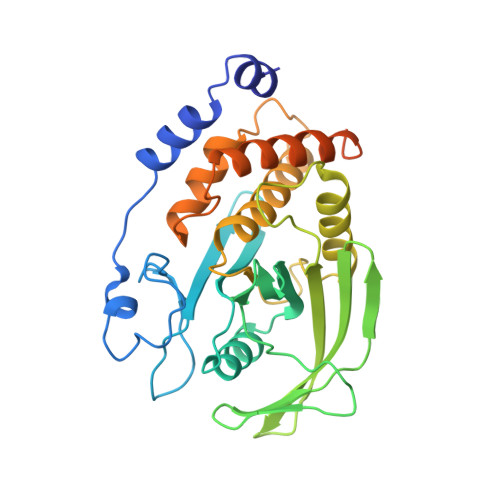
    Protein tyrosine phosphatases (PTPs) constitute a family of receptor-like and cytoplasmic signal transducing enzymes that catalyze the dephosphorylation of phosphotyrosine residues and are characterized by homologous catalytic domains. The crystal structure of a representative member of this family, the 37-kilodalton form (residues 1 to 321) of PTP1B, has been determined at 2.8 A resolution. The enzyme consists of a single domain with the catalytic site located at the base of a shallow cleft. The phosphate recognition site is created from a loop that is located at the amino-terminus of an alpha helix. This site is formed from an 11-residue sequence motif that is diagnostic of PTPs and the dual specificity phosphatases, and that contains the catalytically essential cysteine and arginine residues. The position of the invariant cysteine residue within the phosphate binding site is consistent with its role as a nucleophile in the catalytic reaction. The structure of PTP1B should serve as a model for other members of the PTP family and as a framework for understanding the mechanism of tyrosine dephosphorylation.


  • Organizational Affiliation

    W.M. Keck Structural Biology Laboratory, Cold Spring Harbor Laboratory, NY 11724.


Macromolecules
Find similar proteins by:  (by identity cutoff)  |  3D Structure
Entity ID: 1
MoleculeChains Sequence LengthOrganismDetailsImage
PROTEIN-TYROSINE PHOSPHATASE-1B321Homo sapiensMutation(s): 0 
EC: 3.1.3.48
UniProt & NIH Common Fund Data Resources
Find proteins for P18031 (Homo sapiens)
Explore P18031 
Go to UniProtKB:  P18031
PHAROS:  P18031
GTEx:  ENSG00000196396 
Entity Groups  
Sequence Clusters30% Identity50% Identity70% Identity90% Identity95% Identity100% Identity
UniProt GroupP18031
Sequence Annotations
Expand
  • Reference Sequence
Experimental Data & Validation

Experimental Data

  • Method: X-RAY DIFFRACTION
  • Resolution: 2.85 Å
  • R-Value Work: 0.208 
  • R-Value Observed: 0.208 
  • Space Group: P 31 2 1
Unit Cell:
Length ( Å )Angle ( ˚ )
a = 88.3α = 90
b = 88.3β = 90
c = 104γ = 120
Software Package:
Software NamePurpose
X-PLORmodel building
X-PLORrefinement
X-PLORphasing

Structure Validation

View Full Validation Report


Entry History 

Deposition Data

Revision History  (Full details and data files)

  • Version 1.0: 1994-12-20
    Type: Initial release
  • Version 1.1: 2008-03-24
    Changes: Version format compliance
  • Version 1.2: 2011-07-13
    Changes: Version format compliance
  • Version 1.3: 2017-11-29
    Changes: Derived calculations, Other
  • Version 1.4: 2024-02-14
    Changes: Data collection, Database references