2HNH | pdb_00002hnh

Crystal structure of the catalytic alpha subunit of E. coli replicative DNA polymerase III


Experimental Data Snapshot

  • Method: X-RAY DIFFRACTION
  • Resolution: 2.30 Å
  • R-Value Free: 
    0.258 (Depositor), 0.253 (DCC) 
  • R-Value Work: 
    0.186 (Depositor), 0.184 (DCC) 
  • R-Value Observed: 
    0.190 (Depositor) 

wwPDB Validation 3D Report Full Report

Validation slider image for 2HNH

This is version 1.3 of the entry. See complete history

Literature

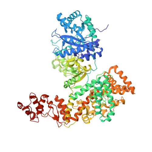
Crystal Structure of the Catalytic alpha Subunit of E. coli Replicative DNA Polymerase III.

Lamers, M.H.Georgescu, R.E.Lee, S.G.O'donnell, M.Kuriyan, J.

(2006) Cell 126: 881-892

  • DOI: https://doi.org/10.1016/j.cell.2006.07.028
  • Primary Citation Related Structures: 
    2HNH, 2HQA

  • PubMed Abstract: 

    Bacterial replicative DNA polymerases such as Polymerase III (Pol III) share no sequence similarity with other polymerases. The crystal structure, determined at 2.3 A resolution, of a large fragment of Pol III (residues 1-917), reveals a unique chain fold with localized similarity in the catalytic domain to DNA polymerase beta and related nucleotidyltransferases. The structure of Pol III is strikingly different from those of members of the canonical DNA polymerase families, which include eukaryotic replicative polymerases, suggesting that the DNA replication machinery in bacteria arose independently. A structural element near the active site in Pol III that is not present in nucleotidyltransferases but which resembles an element at the active sites of some canonical DNA polymerases suggests that, at a more distant level, all DNA polymerases may share a common ancestor. The structure also suggests a model for interaction of Pol III with the sliding clamp and DNA.


  • Organizational Affiliation
    • Howard Hughes Medical Institute, Department of Molecular and Cell Biology and Department of Chemistry, University of California, Berkeley, CA 94720, USA.

Macromolecule Content 

  • Total Structure Weight: 102.05 kDa 
  • Atom Count: 7,621 
  • Modeled Residue Count: 910 
  • Deposited Residue Count: 910 
  • Unique protein chains: 1

Macromolecules

Find similar proteins by:|  3D Structure
Entity ID: 1
MoleculeChains  Sequence LengthOrganismDetailsImage
DNA polymerase III alpha subunit910Escherichia coliMutation(s): 0 
Gene Names: dnaEpolC
EC: 2.7.7.7
UniProt
Find proteins for P10443 (Escherichia coli (strain K12))
Explore P10443 
Go to UniProtKB:  P10443
Entity Groups
Sequence Clusters30% Identity50% Identity70% Identity90% Identity95% Identity100% Identity
UniProt GroupP10443
Sequence Annotations
Expand
Reference Sequence

Experimental Data & Validation

Experimental Data

  • Method: X-RAY DIFFRACTION
  • Resolution: 2.30 Å
  • R-Value Free:  0.258 (Depositor), 0.253 (DCC) 
  • R-Value Work:  0.186 (Depositor), 0.184 (DCC) 
  • R-Value Observed: 0.190 (Depositor) 
Space Group: P 21 21 21
Unit Cell:
Length ( Å )Angle ( ˚ )
a = 82.584α = 90
b = 92.6β = 90
c = 130.235γ = 90
Software Package:
Software NamePurpose
SCALAdata scaling
SHELXphasing
RESOLVEphasing
REFMACrefinement
PDB_EXTRACTdata extraction
MOSFLMdata reduction
CCP4data scaling
SHELXDphasing

Structure Validation

View Full Validation Report


Entry History 

Deposition Data

Revision History  (Full details and data files)

  • Version 1.0: 2006-09-19
    Type: Initial release
  • Version 1.1: 2008-05-01
    Changes: Version format compliance
  • Version 1.2: 2011-07-13
    Changes: Advisory, Version format compliance
  • Version 1.3: 2024-02-14
    Changes: Data collection, Database references, Derived calculations