2HMZ | pdb_00002hmz

THE STRUCTURES OF MET AND AZIDOMET HEMERYTHRIN AT 1.66 ANGSTROMS RESOLUTION


Experimental Data Snapshot

  • Method: X-RAY DIFFRACTION
  • Resolution: 1.66 Å
  • R-Value Work: 
    0.162 (DCC) 
  • R-Value Observed: 
    0.176 (Depositor) 

wwPDB Validation 3D Report Full Report

Validation slider image for 2HMZ

This is version 1.3 of the entry. See complete history

Literature

Structures of met and azidomet hemerythrin at 1.66 A resolution.

Holmes, M.A.Stenkamp, R.E.

(1991) J Mol Biology 220: 723-737

  • DOI: https://doi.org/10.1016/0022-2836(91)90113-k
  • Primary Citation Related Structures: 
    2HMQ, 2HMZ

  • PubMed Abstract: 

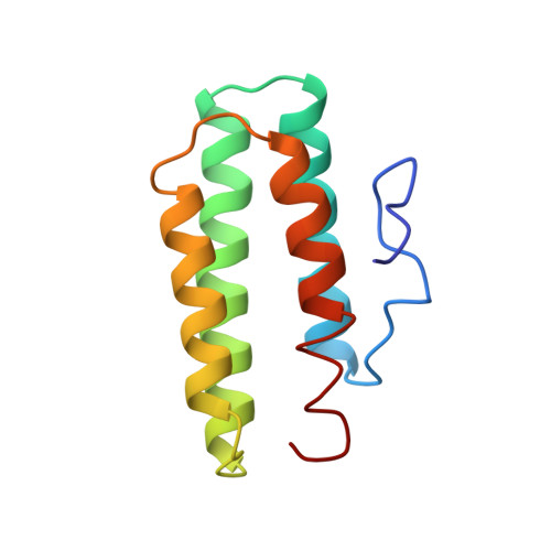
    The crystallographic refinement of met and azidomet hemerythrin has been carried out at 1.66 A resolution in an attempt to characterize precisely the binuclear iron center in this protein. Restrained least-squares refinement has produced molecular models giving R-values of 18.9% for met (65,683 reflections from 10 A to 1.66 A) and 17.6% for azidomet hemerythrin (68,747 reflections from 10.0 A to 1.66 A). The protein structure in each derivative is very similar to that of myohemerythrin. The mu-oxo bridged iron center differs between the two forms. The complex in met hemerythrin is asymmetric with the bridging oxygen closer to one of the iron atoms while the complex in azidomet hemerythrin is symmetric. After investigations of the effects of correlation in the refinement, we believe this difference between the two complexes is associated with chemical differences and is not a refinement artefact.


  • Organizational Affiliation
    • Department of Biological Structure, University of Washington, Seattle 98195.

Macromolecule Content 

  • Total Structure Weight: 54.33 kDa 
  • Atom Count: 4,151 
  • Modeled Residue Count: 456 
  • Deposited Residue Count: 456 
  • Unique protein chains: 1

Macromolecules

Find similar proteins by:|  3D Structure
Entity ID: 1
MoleculeChains  Sequence LengthOrganismDetailsImage
HEMERYTHRIN
A, B, C, D
113Themiste dyscritumMutation(s): 1 
UniProt
Find proteins for P02246 (Themiste dyscrita)
Explore P02246 
Go to UniProtKB:  P02246
Entity Groups
Sequence Clusters30% Identity50% Identity70% Identity90% Identity95% Identity100% Identity
UniProt GroupP02246
Sequence Annotations
Expand
Reference Sequence

Small Molecules

Ligands 2 Unique
IDChains Name / Formula / InChI Key2D Diagram3D Interactions
FEA

Query on FEA


Download:Ideal Coordinates CCD File
F [auth A],
H [auth B],
J [auth C],
L [auth D]
MONOAZIDO-MU-OXO-DIIRON
Fe2 N3 O
GTUZFHRZTDZJHZ-UHFFFAOYSA-N
ACT

Query on ACT


Download:Ideal Coordinates CCD File
E [auth A],
G [auth B],
I [auth C],
K [auth D]
ACETATE ION
C2 H3 O2
QTBSBXVTEAMEQO-UHFFFAOYSA-M

Experimental Data & Validation

Experimental Data

  • Method: X-RAY DIFFRACTION
  • Resolution: 1.66 Å
  • R-Value Work:  0.162 (DCC) 
  • R-Value Observed: 0.176 (Depositor) 
Space Group: P 4
Unit Cell:
Length ( Å )Angle ( ˚ )
a = 86.6α = 90
b = 86.6β = 90
c = 80.8γ = 90
Software Package:
Software NamePurpose
PROFFTrefinement

Structure Validation

View Full Validation Report


Entry History 

Deposition Data

Revision History  (Full details and data files)

  • Version 1.0: 1992-01-15
    Type: Initial release
  • Version 1.1: 2008-03-03
    Changes: Version format compliance
  • Version 1.2: 2011-07-13
    Changes: Derived calculations, Version format compliance
  • Version 1.3: 2024-06-05
    Changes: Data collection, Database references, Derived calculations, Other