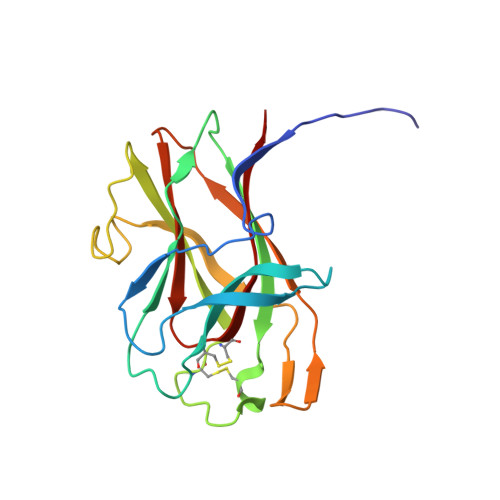
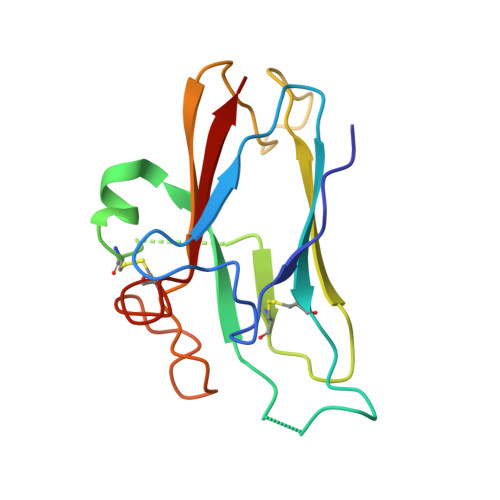
2HLE

Structural and biophysical characterization of the EPHB4-EPHRINB2 protein protein interaction and receptor specificity.


Experimental Data Snapshot

  • Method: X-RAY DIFFRACTION
  • Resolution: 2.05 Å
  • R-Value Free: 0.295 
  • R-Value Work: 0.226 
  • R-Value Observed: 0.229 

Starting Model: experimental
View more details

wwPDB Validation   3D Report Full Report


This is version 1.3 of the entry. See complete history


Literature

Structural and Biophysical Characterization of the EphB4-EphrinB2 Protein-Protein Interaction and Receptor Specificity.

Chrencik, J.E.Brooun, A.Kraus, M.L.Recht, M.I.Kolatkar, A.R.Han, G.W.Seifert, J.M.Widmer, H.Auer, M.Kuhn, P.

(2006) J Biol Chem 281: 28185-28192

  • DOI: https://doi.org/10.1074/jbc.M605766200
  • Primary Citation of Related Structures:  
    2HLE

  • PubMed Abstract: 

    Increasing evidence implicates the interaction of the EphB4 receptor with its preferred ligand, ephrinB2, in pathological forms of angiogenesis and in tumorigenesis. To identify the molecular determinants of the unique specificity of EphB4 for ephrinB2, we determined the crystal structure of the ligand binding domain of EphB4 in complex with the extracellular domain of ephrinB2. This structural analysis suggested that one amino acid, Leu-95, plays a particularly important role in defining the structural features that confer the ligand selectivity of EphB4. Indeed, all other Eph receptors, which promiscuously bind many ephrins, have a conserved arginine at the position corresponding to Leu-95 of EphB4. We have also found that amino acid changes in the EphB4 ligand binding cavity, designed based on comparison with the crystal structure of the more promiscuous EphB2 receptor, yield EphB4 variants with altered binding affinity for ephrinB2 and an antagonistic peptide. Isothermal titration calorimetry experiments with an EphB4 Leu-95 to arginine mutant confirmed the importance of this amino acid in conferring high affinity binding to both ephrinB2 and the antagonistic peptide ligand. Isothermal titration calorimetry measurements also revealed an interesting thermodynamic discrepancy between ephrinB2 binding, which is an entropically driven process, and peptide binding, which is an enthalpically driven process. These results provide critical information on the EphB4*ephrinB2 protein interfaces and their mode of interaction, which will facilitate development of small molecule compounds inhibiting the EphB4*ephrinB2 interaction as novel cancer therapeutics.


  • Organizational Affiliation

    Department of Cellular Biology, The Scripps Research Institute, La Jolla, California 92037, USA.


Macromolecules
Find similar proteins by:  (by identity cutoff)  |  3D Structure
Entity ID: 1
MoleculeChains Sequence LengthOrganismDetailsImage
Ephrin type-B receptor 4188Homo sapiensMutation(s): 0 
Gene Names: EPHB4HTK
EC: 2.7.10.1
UniProt & NIH Common Fund Data Resources
Find proteins for P54760 (Homo sapiens)
Explore P54760 
Go to UniProtKB:  P54760
PHAROS:  P54760
GTEx:  ENSG00000196411 
Entity Groups  
Sequence Clusters30% Identity50% Identity70% Identity90% Identity95% Identity100% Identity
UniProt GroupP54760
Sequence Annotations
Expand
  • Reference Sequence
Find similar proteins by:  (by identity cutoff)  |  3D Structure
Entity ID: 2
MoleculeChains Sequence LengthOrganismDetailsImage
Ephrin-B2138Homo sapiensMutation(s): 0 
Gene Names: EFNB2EPLG5HTKLLERK5
UniProt & NIH Common Fund Data Resources
Find proteins for P52799 (Homo sapiens)
Explore P52799 
Go to UniProtKB:  P52799
PHAROS:  P52799
GTEx:  ENSG00000125266 
Entity Groups  
Sequence Clusters30% Identity50% Identity70% Identity90% Identity95% Identity100% Identity
UniProt GroupP52799
Sequence Annotations
Expand
  • Reference Sequence
Experimental Data & Validation

Experimental Data

  • Method: X-RAY DIFFRACTION
  • Resolution: 2.05 Å
  • R-Value Free: 0.295 
  • R-Value Work: 0.226 
  • R-Value Observed: 0.229 
  • Space Group: P 41
Unit Cell:
Length ( Å )Angle ( ˚ )
a = 81.085α = 90
b = 81.085β = 90
c = 50.945γ = 90
Software Package:
Software NamePurpose
REFMACrefinement
HKL-2000data reduction
HKL-2000data scaling
MOLREPphasing

Structure Validation

View Full Validation Report


Entry History 

Deposition Data

Revision History  (Full details and data files)

  • Version 1.0: 2006-08-22
    Type: Initial release
  • Version 1.1: 2008-05-01
    Changes: Version format compliance
  • Version 1.2: 2011-07-13
    Changes: Advisory, Version format compliance
  • Version 1.3: 2023-08-30
    Changes: Data collection, Database references, Refinement description