2HLA

SPECIFICITY POCKETS FOR THE SIDE CHAINS OF PEPTIDE ANTIGENS IN HLA-AW68


Experimental Data Snapshot

  • Method: X-RAY DIFFRACTION
  • Resolution: 2.60 Å
  • R-Value Work: 0.173 

wwPDB Validation   3D Report Full Report


This is version 1.3 of the entry. See complete history


Literature

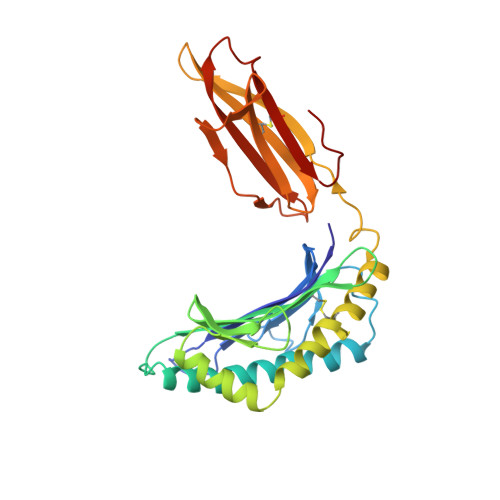
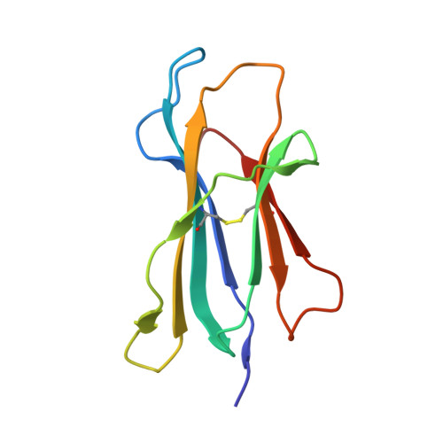
Specificity pockets for the side chains of peptide antigens in HLA-Aw68.

Garrett, T.P.Saper, M.A.Bjorkman, P.J.Strominger, J.L.Wiley, D.C.

(1989) Nature 342: 692-696

  • DOI: https://doi.org/10.1038/342692a0
  • Primary Citation of Related Structures:  
    2HLA

  • PubMed Abstract: 

    We have determined the structure of a second human histocompatibility glycoprotein, HLA-Aw68, by X-ray crystallography and refined it to a resolution of 2.6 A. Overall, the structure is extremely similar to that of HLA-A2 (refs 1, 2; and M.A.S. et al., manuscript in preparation), although the 11 amino-acid substitutions at polymorphic residues in the antigen-binding cleft alter the detailed shape and electrostatic charge of that site. A prominent negatively charged pocket within the cleft extends underneath the alpha-helix of the alpha 1-domain, providing a potential subsite for recognizing a positively charged side chain or peptide N terminus. Uninterpreted electron density, presumably representing an unknown 'antigen(s)', which seems to be different from that seen in the HLA-A2 structure, occupies the cleft and extends into the negatively charged pocket in HLA-Aw68. The structures of HLA-Aw68 and HLA-A2 demonstrate how polymorphism creates and alters subsites (pockets) positioned to bind peptide side chains, thereby suggesting the structural basis for allelic specificity in foreign antigen binding.


  • Organizational Affiliation

    Department of Biochemistry and Molecular Biology, Harvard University, Cambridge, Massachusetts 02138.


Macromolecules
Find similar proteins by:  (by identity cutoff)  |  3D Structure
Entity ID: 1
MoleculeChains Sequence LengthOrganismDetailsImage
CLASS I HISTOCOMPATIBILITY ANTIGEN (HLA-Aw68)270Homo sapiensMutation(s): 0 
UniProt & NIH Common Fund Data Resources
Find proteins for P04439 (Homo sapiens)
Explore P04439 
Go to UniProtKB:  P04439
PHAROS:  P04439
GTEx:  ENSG00000206503 
Entity Groups  
Sequence Clusters30% Identity50% Identity70% Identity90% Identity95% Identity100% Identity
UniProt GroupP04439
Sequence Annotations
Expand
  • Reference Sequence
Find similar proteins by:  (by identity cutoff)  |  3D Structure
Entity ID: 2
MoleculeChains Sequence LengthOrganismDetailsImage
BETA 2-MICROGLOBULIN99Homo sapiensMutation(s): 0 
UniProt & NIH Common Fund Data Resources
Find proteins for P61769 (Homo sapiens)
Explore P61769 
Go to UniProtKB:  P61769
PHAROS:  P61769
GTEx:  ENSG00000166710 
Entity Groups  
Sequence Clusters30% Identity50% Identity70% Identity90% Identity95% Identity100% Identity
UniProt GroupP61769
Sequence Annotations
Expand
  • Reference Sequence
Experimental Data & Validation

Experimental Data

  • Method: X-RAY DIFFRACTION
  • Resolution: 2.60 Å
  • R-Value Work: 0.173 
  • Space Group: P 21 21 21
Unit Cell:
Length ( Å )Angle ( ˚ )
a = 60α = 90
b = 80.66β = 90
c = 113γ = 90
Software Package:
Software NamePurpose
X-PLORrefinement

Structure Validation

View Full Validation Report


Entry History 

Deposition Data

Revision History  (Full details and data files)

  • Version 1.0: 1990-04-15
    Type: Initial release
  • Version 1.1: 2008-03-24
    Changes: Version format compliance
  • Version 1.2: 2011-07-13
    Changes: Version format compliance
  • Version 1.3: 2017-11-29
    Changes: Derived calculations, Other