2HJG

The crystal structure of the B. subtilis YphC GTPase in complex with GDP


Experimental Data Snapshot

  • Method: X-RAY DIFFRACTION
  • Resolution: 2.50 Å
  • R-Value Free: 0.274 
  • R-Value Work: 0.215 
  • R-Value Observed: 0.218 

wwPDB Validation   3D Report Full Report


Ligand Structure Quality Assessment 


This is version 1.3 of the entry. See complete history


Literature

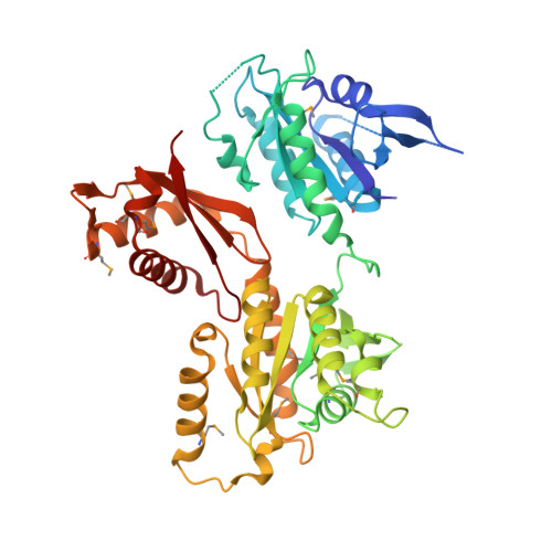
The essential GTPase YphC displays a major domain rearrangement associated with nucleotide binding.

Muench, S.P.Xu, L.Sedelnikova, S.E.Rice, D.W.

(2006) Proc Natl Acad Sci U S A 103: 12359-12364

  • DOI: https://doi.org/10.1073/pnas.0602585103
  • Primary Citation of Related Structures:  
    2HJG

  • PubMed Abstract: 

    The structure of a Bacillus subtilis YphC/GDP complex shows that it contains two GTPase domains that pack against a central domain whose fold resembles that of an RNA binding KH-domain. Comparisons of this structure to that of a homologue in Thermotoga maritima reveals a dramatic rearrangement in the position of the N-terminal GTPase domain with a shift of up to 60 A and the formation of a totally different interface to the central domain. This rearrangement appears to be triggered by conformational changes of the switch II region in this domain in response to nucleotide binding. Modeling studies suggest that this motion represents transitions between the "on" and "off" states of the GTPase, the effect of which is to alternately expose and bury a positively charged face of the central domain that we suggest is involved in RNA recognition as part of the possible role of this enzyme in ribosome binding.


  • Organizational Affiliation

    Department of Molecular Biology and Biotechnology, University of Sheffield, Sheffield S10 2TN, United Kingdom.


Macromolecules
Find similar proteins by:  (by identity cutoff)  |  3D Structure
Entity ID: 1
MoleculeChains Sequence LengthOrganismDetailsImage
GTP-binding protein engA436Bacillus subtilisMutation(s): 12 
Gene Names: engA
UniProt
Find proteins for P50743 (Bacillus subtilis (strain 168))
Explore P50743 
Go to UniProtKB:  P50743
Entity Groups  
Sequence Clusters30% Identity50% Identity70% Identity90% Identity95% Identity100% Identity
UniProt GroupP50743
Sequence Annotations
Expand
  • Reference Sequence
Small Molecules
Modified Residues  1 Unique
IDChains TypeFormula2D DiagramParent
MSE
Query on MSE
A
L-PEPTIDE LINKINGC5 H11 N O2 SeMET
Experimental Data & Validation

Experimental Data

  • Method: X-RAY DIFFRACTION
  • Resolution: 2.50 Å
  • R-Value Free: 0.274 
  • R-Value Work: 0.215 
  • R-Value Observed: 0.218 
  • Space Group: P 21 21 21
Unit Cell:
Length ( Å )Angle ( ˚ )
a = 62.711α = 90
b = 65.045β = 90
c = 110.61γ = 90
Software Package:
Software NamePurpose
REFMACrefinement
MOSFLMdata reduction
CCP4data scaling
SOLVEphasing

Structure Validation

View Full Validation Report


Ligand Structure Quality Assessment 


Entry History 

Deposition Data

Revision History  (Full details and data files)

  • Version 1.0: 2006-08-08
    Type: Initial release
  • Version 1.1: 2008-05-01
    Changes: Version format compliance
  • Version 1.2: 2011-07-13
    Changes: Version format compliance
  • Version 1.3: 2018-01-24
    Changes: Structure summary