2HJ4

Crystal structure of Alcaligenes faecalis AADH complex with p-nitrobenzylamine


Experimental Data Snapshot

  • Method: X-RAY DIFFRACTION
  • Resolution: 1.80 Å
  • R-Value Free: 0.205 
  • R-Value Work: 0.159 
  • R-Value Observed: 0.161 

wwPDB Validation   3D Report Full Report


Ligand Structure Quality Assessment 


This is version 1.1 of the entry. See complete history


Literature

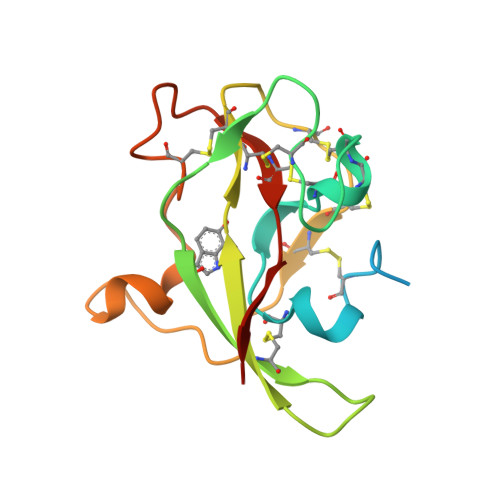
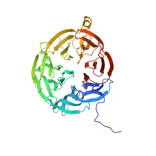
Isotope effects reveal that para-substituted benzylamines are poor reactivity probes of the quinoprotein mechanism for aromatic amine dehydrogenase.

Hothi, P.Roujeinikova, A.Khadra, K.A.Lee, M.Cullis, P.Leys, D.Scrutton, N.S.

(2007) Biochemistry 46: 9250-9259

  • DOI: https://doi.org/10.1021/bi7007239
  • Primary Citation of Related Structures:  
    2HJ4, 2HJB, 2Q7Q

  • PubMed Abstract: 

    Structure-activity correlations have been employed previously in the mechanistic interpretation of TTQ-dependent amine dehydrogenases using a series of para-substituted benzylamines. However, by combining the use of kinetic isotope effects (KIEs) and crystallographic analysis, in conjunction with structure-reactivity correlation studies, we show that para-substituted benzylamines are poor reactivity probes for TTQ-dependent aromatic amine dehydrogenase (AADH). Stopped-flow kinetic studies of the reductive half-reaction, with para-substituted benzylamines and their dideuterated counterparts, demonstrate that C-H or C-D bond breakage is not fully rate limiting (KIEs approximately unity). Contrary to previous reports, Hammett plots exhibit a poor correlation of structure-reactivity data with electronic substituent effects for para-substituted benzylamines and phenylethylamines. Crystallographic studies of enzyme-substrate complexes reveal that the observed structure-reactivity correlations are not attributed to distinct binding modes for para-substituted benzylamines in the active site, although two binding sites for p-nitrobenzylamine are identified. We identify structural rearrangements, prior to the H-transfer step, which are likely to limit the rate of TTQ reduction by benzylamines. This work emphasizes (i) the need for caution when applying structure-activity correlations to enzyme-catalyzed reactions and (ii) the added benefit of using both isotope effects and structural analysis, in conjunction with structure-reactivity relationships, to study chemical steps in enzyme reaction cycles.


  • Organizational Affiliation

    Manchester Interdisciplinary Biocentre, Faculty of Life Sciences, University of Manchester, UK.


Macromolecules
Find similar proteins by:  (by identity cutoff)  |  3D Structure
Entity ID: 1
MoleculeChains Sequence LengthOrganismDetailsImage
Aromatic amine dehydrogenase; chain D, HA [auth D],
B [auth H]
135Alcaligenes faecalisMutation(s): 0 
EC: 1.4.99.4
UniProt
Find proteins for P84887 (Alcaligenes faecalis)
Explore P84887 
Go to UniProtKB:  P84887
Entity Groups  
Sequence Clusters30% Identity50% Identity70% Identity90% Identity95% Identity100% Identity
UniProt GroupP84887
Sequence Annotations
Expand
  • Reference Sequence
Find similar proteins by:  (by identity cutoff)  |  3D Structure
Entity ID: 2
MoleculeChains Sequence LengthOrganismDetailsImage
Aromatic amine dehydrogenase; chain A, BC [auth A],
D [auth B]
361Alcaligenes faecalisMutation(s): 0 
EC: 1.4.99.4
UniProt
Find proteins for P84888 (Alcaligenes faecalis)
Explore P84888 
Go to UniProtKB:  P84888
Entity Groups  
Sequence Clusters30% Identity50% Identity70% Identity90% Identity95% Identity100% Identity
UniProt GroupP84888
Sequence Annotations
Expand
  • Reference Sequence
Small Molecules
Modified Residues  1 Unique
IDChains TypeFormula2D DiagramParent
TRQ
Query on TRQ
A [auth D],
B [auth H]
L-PEPTIDE LINKINGC11 H10 N2 O4TRP
Binding Affinity Annotations 
IDSourceBinding Affinity
PNZ Binding MOAD:  2HJ4 Kd: 4290 (nM) from 1 assay(s)
PDBBind:  2HJ4 Kd: 4290 (nM) from 1 assay(s)
Experimental Data & Validation

Experimental Data

  • Method: X-RAY DIFFRACTION
  • Resolution: 1.80 Å
  • R-Value Free: 0.205 
  • R-Value Work: 0.159 
  • R-Value Observed: 0.161 
  • Space Group: P 21 21 21
Unit Cell:
Length ( Å )Angle ( ˚ )
a = 91.254α = 90
b = 96.544β = 90
c = 120.632γ = 90
Software Package:
Software NamePurpose
REFMACrefinement
PDB_EXTRACTdata extraction
MOSFLMdata reduction
SCALAdata scaling

Structure Validation

View Full Validation Report


Ligand Structure Quality Assessment 


Entry History 

Deposition Data

Revision History  (Full details and data files)

  • Version 1.0: 2007-11-06
    Type: Initial release
  • Version 1.1: 2011-07-13
    Changes: Derived calculations, Version format compliance