2HEK | pdb_00002hek

Crystal structure of O67745, a hypothetical protein from Aquifex aeolicus at 2.0 A resolution.


Experimental Data Snapshot

  • Method: X-RAY DIFFRACTION
  • Resolution: 2.00 Å
  • R-Value Free: 
    0.220 (Depositor), 0.237 (DCC) 
  • R-Value Work: 
    0.175 (Depositor), 0.197 (DCC) 
  • R-Value Observed: 
    0.177 (Depositor) 

wwPDB Validation 3D Report Full Report

Validation slider image for 2HEK

Ligand Structure Quality Assessment 


This is version 1.4 of the entry. See complete history

Literature

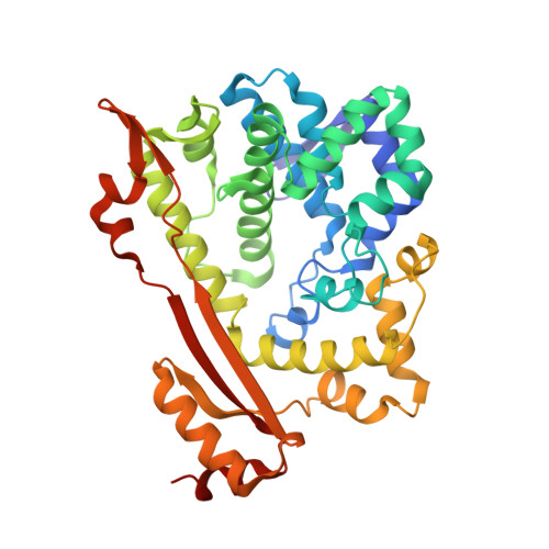
Structure of O67745_AQUAE, a hypothetical protein from Aquifex aeolicus.

Oganesyan, V.Adams, P.D.Jancarik, J.Kim, R.Kim, S.H.

(2007) Acta Crystallogr Sect F Struct Biol Cryst Commun 63: 369-374

  • DOI: https://doi.org/10.1107/S1744309107018945
  • Primary Citation Related Structures: 
    2HEK

  • PubMed Abstract: 

    Using single-wavelength anomalous dispersion data obtained from a gold-derivatized crystal, the X-ray crystal structure of the protein 067745_AQUAE from the prokaryotic organism Aquifex aeolicus has been determined to a resolution of 2.0 A. Amino-acid residues 1-371 of the 44 kDa protein were identified by Pfam as an HD domain and a member of the metal-dependent phosphohydrolase superfamily (accession No. PF01966). Although three families from this large and diverse group of enzymatic proteins are represented in the PDB, the structure of 067745_AQUAE reveals a unique fold that is unlike the others and that is likely to represent a new subfamily, further organizing the families and characterizing the proteins. Data are presented that provide the first insights into the structural organization of the proteins within this clan and a distal alternative GDP-binding domain outside the metal-binding active site is proposed.


  • Organizational Affiliation
    • Berkeley Structural Genomics Center, Physical Biosciences Division, Lawrence Berkeley National Laboratory, Berkeley, California 94720, USA.

Macromolecule Content 

  • Total Structure Weight: 90.76 kDa 
  • Atom Count: 6,705 
  • Modeled Residue Count: 738 
  • Deposited Residue Count: 742 
  • Unique protein chains: 1

Macromolecules

Find similar proteins by:|  3D Structure
Entity ID: 1
MoleculeChains  Sequence LengthOrganismDetailsImage
Hypothetical protein
A, B
371Aquifex aeolicusMutation(s): 0 
UniProt
Find proteins for O67745 (Aquifex aeolicus (strain VF5))
Explore O67745 
Go to UniProtKB:  O67745
Entity Groups
Sequence Clusters30% Identity50% Identity70% Identity90% Identity95% Identity100% Identity
UniProt GroupO67745
Sequence Annotations
Expand
Reference Sequence

Small Molecules

Ligands 6 Unique
IDChains Name / Formula / InChI Key2D Diagram3D Interactions
GDP

Query on GDP


Download:Ideal Coordinates CCD File
G [auth A],
H [auth A]
GUANOSINE-5'-DIPHOSPHATE
C10 H15 N5 O11 P2
QGWNDRXFNXRZMB-UUOKFMHZSA-N
PO4

Query on PO4


Download:Ideal Coordinates CCD File
C [auth A],
O [auth B],
P [auth B],
Q [auth B]
PHOSPHATE ION
O4 P
NBIIXXVUZAFLBC-UHFFFAOYSA-K
GOL

Query on GOL


Download:Ideal Coordinates CCD File
I [auth A]
J [auth A]
K [auth A]
L [auth A]
M [auth A]
I [auth A],
J [auth A],
K [auth A],
L [auth A],
M [auth A],
N [auth A],
W [auth B],
X [auth B]
GLYCEROL
C3 H8 O3
PEDCQBHIVMGVHV-UHFFFAOYSA-N
BR

Query on BR


Download:Ideal Coordinates CCD File
S [auth B]BROMIDE ION
Br
CPELXLSAUQHCOX-UHFFFAOYSA-M
ZN

Query on ZN


Download:Ideal Coordinates CCD File
D [auth A],
R [auth B]
ZINC ION
Zn
PTFCDOFLOPIGGS-UHFFFAOYSA-N
CL

Query on CL


Download:Ideal Coordinates CCD File
E [auth A],
F [auth A],
T [auth B],
U [auth B],
V [auth B]
CHLORIDE ION
Cl
VEXZGXHMUGYJMC-UHFFFAOYSA-M

Experimental Data & Validation

Experimental Data

  • Method: X-RAY DIFFRACTION
  • Resolution: 2.00 Å
  • R-Value Free:  0.220 (Depositor), 0.237 (DCC) 
  • R-Value Work:  0.175 (Depositor), 0.197 (DCC) 
  • R-Value Observed: 0.177 (Depositor) 
Space Group: C 2 2 21
Unit Cell:
Length ( Å )Angle ( ˚ )
a = 107.687α = 90
b = 144.413β = 90
c = 144.677γ = 90
Software Package:
Software NamePurpose
REFMACrefinement
PHENIXphasing

Structure Validation

View Full Validation Report


Ligand Structure Quality Assessment 


Entry History 

Deposition Data

Revision History  (Full details and data files)

  • Version 1.0: 2006-07-04
    Type: Initial release
  • Version 1.1: 2008-02-06
    Changes: Version format compliance
  • Version 1.2: 2011-07-13
    Changes: Derived calculations, Version format compliance
  • Version 1.3: 2017-10-18
    Changes: Refinement description
  • Version 1.4: 2024-02-14
    Changes: Data collection, Database references, Derived calculations