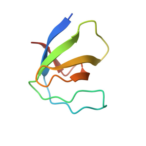
2HDA

Yes SH3 domain


Experimental Data Snapshot

  • Method: X-RAY DIFFRACTION
  • Resolution: 1.90 Å
  • R-Value Free: 0.270 
  • R-Value Work: 0.214 
  • R-Value Observed: 0.216 

Starting Model: experimental
View more details

wwPDB Validation   3D Report Full Report


This is version 1.3 of the entry. See complete history


Literature

Crystallographic structure of the SH3 domain of the human c-Yes tyrosine kinase: Loop flexibility and amyloid aggregation.

Martin-Garcia, J.M.Luque, I.Mateo, P.L.Ruiz-Sanz, J.Camara-Artigas, A.

(2007) FEBS Lett 581: 1701-1706

  • DOI: https://doi.org/10.1016/j.febslet.2007.03.059
  • Primary Citation of Related Structures:  
    2HDA

  • PubMed Abstract: 

    SH3 domains from the Src family of tyrosine kinases represent an interesting example of the delicate balance between promiscuity and specificity characteristic of proline-rich ligand recognition by SH3 domains. The development of inhibitors of therapeutic potential requires a good understanding of the molecular determinants of binding affinity and specificity and relies on the availability of high quality structural information. Here, we present the first high-resolution crystal structure of the SH3 domain of the c-Yes oncogen. Comparison with other SH3 domains from the Src family revealed significant deviations in the loop regions. In particular, the n-Src loop, highly flexible and partially disordered, is stabilized in an unusual conformation by the establishment of several intramolecular hydrogen bonds. Additionally, we present here the first report of amyloid aggregation by an SH3 domain from the Src family.


  • Organizational Affiliation

    Department of Physical Chemistry and Institute of Biotechnology, Faculty of Sciences, University of Granada, 18071 Granada, Spain.


Macromolecules
Find similar proteins by:  (by identity cutoff)  |  3D Structure
Entity ID: 1
MoleculeChains Sequence LengthOrganismDetailsImage
Proto-oncogene tyrosine-protein kinase Yes64Homo sapiensMutation(s): 0 
Gene Names: YES1YES
EC: 2.7.10.2
UniProt & NIH Common Fund Data Resources
Find proteins for P07947 (Homo sapiens)
Explore P07947 
Go to UniProtKB:  P07947
PHAROS:  P07947
GTEx:  ENSG00000176105 
Entity Groups  
Sequence Clusters30% Identity50% Identity70% Identity90% Identity95% Identity100% Identity
UniProt GroupP07947
Sequence Annotations
Expand
  • Reference Sequence
Small Molecules
Ligands 1 Unique
IDChains Name / Formula / InChI Key2D Diagram3D Interactions
SO4
Query on SO4

Download Ideal Coordinates CCD File 
B [auth A]SULFATE ION
O4 S
QAOWNCQODCNURD-UHFFFAOYSA-L
Experimental Data & Validation

Experimental Data

  • Method: X-RAY DIFFRACTION
  • Resolution: 1.90 Å
  • R-Value Free: 0.270 
  • R-Value Work: 0.214 
  • R-Value Observed: 0.216 
  • Space Group: P 63 2 2
Unit Cell:
Length ( Å )Angle ( ˚ )
a = 46.009α = 90
b = 46.009β = 90
c = 90.431γ = 120
Software Package:
Software NamePurpose
REFMACrefinement
MOLREPphasing

Structure Validation

View Full Validation Report


Entry History 

Deposition Data

Revision History  (Full details and data files)

  • Version 1.0: 2007-04-17
    Type: Initial release
  • Version 1.1: 2008-05-01
    Changes: Version format compliance
  • Version 1.2: 2011-07-13
    Changes: Derived calculations, Version format compliance
  • Version 1.3: 2023-08-30
    Changes: Data collection, Database references, Derived calculations, Refinement description