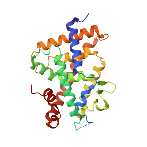
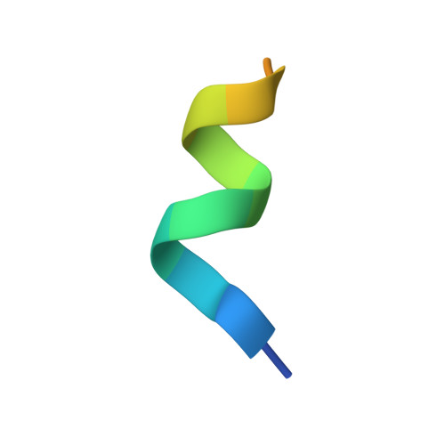
2HCD

Crystal structure of the ligand binding domain of the Vitamin D nuclear receptor in complex with Gemini and a coactivator peptide


Experimental Data Snapshot

  • Method: X-RAY DIFFRACTION
  • Resolution: 2.60 Å
  • R-Value Free: 0.260 
  • R-Value Work: 0.204 
  • R-Value Observed: 0.227 

Starting Model: experimental
View more details

wwPDB Validation   3D Report Full Report


Ligand Structure Quality Assessment 


This is version 1.4 of the entry. See complete history


Literature

Adaptability of the Vitamin D nuclear receptor to the synthetic ligand Gemini: remodelling the LBP with one side chain rotation

Ciesielski, F.Rochel, N.Moras, D.

(2007) J Steroid Biochem Mol Biol 103: 235-242

  • DOI: https://doi.org/10.1016/j.jsbmb.2006.12.003
  • Primary Citation of Related Structures:  
    2HC4, 2HCD

  • PubMed Abstract: 

    The crystal structure of the ligand binding domain (LBD) of the wild-type Vitamin D receptor (VDR) of zebrafish bound to Gemini, a synthetic agonist ligand with two identical side chains branching at carbon 20 reveals a ligand-dependent structural rearrangement of the ligand binding pocket (LBP). The rotation of a Leu side chain opens the access to a channel that can accommodate the second side chain of the ligand. The 25% increase of the LBP's volume does not alter the essential agonist features of VDR. The possibility to adapt the LBP to novel ligands with different chemistry and/or structure opens new perspectives in the design of more specifically targeted ligands.


  • Organizational Affiliation

    Département de Biologie et de Génomique Structurales, IGBMC, CNRS/INSERM Université Louis Pasteur, Parc d'innovation BP10142, 67404 Illkirch Cedex, France.


Macromolecules
Find similar proteins by:  (by identity cutoff)  |  3D Structure
Entity ID: 1
MoleculeChains Sequence LengthOrganismDetailsImage
Vitamin D receptor302Danio rerioMutation(s): 0 
UniProt
Find proteins for Q9PTN2 (Danio rerio)
Explore Q9PTN2 
Go to UniProtKB:  Q9PTN2
Entity Groups  
Sequence Clusters30% Identity50% Identity70% Identity90% Identity95% Identity100% Identity
UniProt GroupQ9PTN2
Sequence Annotations
Expand
  • Reference Sequence

Find similar proteins by:  Sequence   |   3D Structure  

Entity ID: 2
MoleculeChains Sequence LengthOrganismDetailsImage
SRC-1 from Nuclear receptor coactivator 115Homo sapiensMutation(s): 0 
UniProt & NIH Common Fund Data Resources
Find proteins for Q15788 (Homo sapiens)
Explore Q15788 
Go to UniProtKB:  Q15788
PHAROS:  Q15788
GTEx:  ENSG00000084676 
Entity Groups  
Sequence Clusters30% Identity50% Identity70% Identity90% Identity95% Identity100% Identity
UniProt GroupQ15788
Sequence Annotations
Expand
  • Reference Sequence
Small Molecules
Ligands 1 Unique
IDChains Name / Formula / InChI Key2D Diagram3D Interactions
BIV
Query on BIV

Download Ideal Coordinates CCD File 
C [auth A]21-NOR-9,10-SECOCHOLESTA-5,7,10(19)-TRIENE-1,3,25-TRIOL, 20-(4-HYDROXY-4-METHYLPENTYL)-, (1A,3B,5Z,7E)
C32 H54 O4
WTQXZYVWLNPNEX-LKUPKUSPSA-N
Experimental Data & Validation

Experimental Data

  • Method: X-RAY DIFFRACTION
  • Resolution: 2.60 Å
  • R-Value Free: 0.260 
  • R-Value Work: 0.204 
  • R-Value Observed: 0.227 
  • Space Group: P 65 2 2
Unit Cell:
Length ( Å )Angle ( ˚ )
a = 65.859α = 90
b = 65.859β = 90
c = 265.018γ = 120
Software Package:
Software NamePurpose
CNSrefinement
ADSCdata collection
HKL-2000data scaling
CNSphasing

Structure Validation

View Full Validation Report


Ligand Structure Quality Assessment 


Entry History 

Deposition Data

Revision History  (Full details and data files)

  • Version 1.0: 2007-05-01
    Type: Initial release
  • Version 1.1: 2008-05-01
    Changes: Version format compliance
  • Version 1.2: 2011-07-13
    Changes: Version format compliance
  • Version 1.3: 2021-03-03
    Changes: Advisory, Database references, Derived calculations, Source and taxonomy
  • Version 1.4: 2023-10-25
    Changes: Advisory, Data collection, Database references, Refinement description