2HCC | pdb_00002hcc

SOLUTION STRUCTURE OF THE HUMAN CHEMOKINE HCC-2, NMR, 30 STRUCTURES


Experimental Data Snapshot

  • Method: SOLUTION NMR
  • Conformers Calculated: 100 
  • Conformers Submitted: 30 
  • Selection Criteria: ENERGY, AGREEMENT WITH EXPERIMENTAL DATA 

wwPDB Validation 3D Report Full Report

Validation slider image for 2HCC

This is version 1.4 of the entry. See complete history

Literature

Solution structure of the human CC chemokine 2: A monomeric representative of the CC chemokine subtype.

Sticht, H.Escher, S.E.Schweimer, K.Forssmann, W.G.Rosch, P.Adermann, K.

(1999) Biochemistry 38: 5995-6002

  • DOI: https://doi.org/10.1021/bi990065i
  • Primary Citation Related Structures: 
    2HCC

  • PubMed Abstract: 

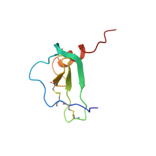
    HCC-2, a 66-amino acid residue human CC chemokine, was reported to induce chemotaxis on monocytes, T-lymphocytes, and eosinophils. The three-dimensional structure of HCC-2 has been determined by 1H nuclear magnetic resonance (NMR) spectroscopy and restrained molecular dynamics calculations on the basis of 871 experimental restraints. The structure is well-defined, exhibiting average root-mean-square deviations of 0.58 and 0.96 A for the backbone heavy atoms and all heavy atoms of residues 5-63, respectively. In contrast to most other chemokines, subtle structural differences impede dimer formation of HCC-2 in a concentration range of 0.1 microM to 2 mM. HCC-2, however, exhibits the same structural elements as the other chemokines, i.e., a triple-stranded antiparallel beta-sheet covered by an alpha-helix, showing that the chemokine fold is not influenced by quaternary interactions. Structural investigations with a HCC-2 mutant prove that a third additional disulfide bond present in wild-type HCC-2 is not necessary for maintaining the relative orientation of the helix and the beta-sheet.


  • Organizational Affiliation
    • Lehrstuhl für Biopolymere, Universität Bayreuth, D-95440 Bayreuth, Germany. Heinrich.Sticht@Uni-Bayreuth.de

Macromolecule Content 

  • Total Structure Weight: 7.22 kDa 
  • Atom Count: 500 
  • Modeled Residue Count: 66 
  • Deposited Residue Count: 66 
  • Unique protein chains: 1

Macromolecules

Find similar proteins by:|  3D Structure
Entity ID: 1
MoleculeChains  Sequence LengthOrganismDetailsImage
HUMAN CHEMOKINE HCC-266Homo sapiensMutation(s): 0 
UniProt & NIH Common Fund Data Resources
Find proteins for Q16663 (Homo sapiens)
Explore Q16663 
Go to UniProtKB:  Q16663
GTEx:  ENSG00000275718 
Entity Groups
Sequence Clusters30% Identity50% Identity70% Identity90% Identity95% Identity100% Identity
UniProt GroupQ16663
Sequence Annotations
Expand
Reference Sequence

Experimental Data & Validation

Experimental Data

  • Method: SOLUTION NMR
  • Conformers Calculated: 100 
  • Conformers Submitted: 30 
  • Selection Criteria: ENERGY, AGREEMENT WITH EXPERIMENTAL DATA 

Structure Validation

View Full Validation Report


Entry History 

Deposition Data

Revision History  (Full details and data files)

  • Version 1.0: 1999-07-13
    Type: Initial release
  • Version 1.1: 2008-03-24
    Changes: Version format compliance
  • Version 1.2: 2011-07-13
    Changes: Version format compliance
  • Version 1.3: 2022-03-09
    Changes: Database references, Derived calculations, Other
  • Version 1.4: 2024-11-06
    Changes: Data collection, Structure summary