2H6E

Crystal structure of the D-arabinose dehydrogenase from Sulfolobus solfataricus


Experimental Data Snapshot

  • Method: X-RAY DIFFRACTION
  • Resolution: 1.80 Å
  • R-Value Free: 0.232 
  • R-Value Work: 0.195 
  • R-Value Observed: 0.197 

wwPDB Validation   3D Report Full Report


This is version 1.6 of the entry. See complete history


Literature

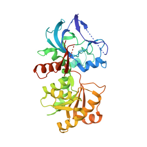
Crystal Structure and Biochemical Properties of the d-Arabinose Dehydrogenase from Sulfolobus solfataricus

Brouns, S.J.Turnbull, A.P.Willemen, H.L.Akerboom, J.van der Oost, J.

(2007) J Mol Biol 371: 1249-1260

  • DOI: https://doi.org/10.1016/j.jmb.2007.05.097
  • Primary Citation of Related Structures:  
    2H6E

  • PubMed Abstract: 

    Sulfolobus solfataricus metabolizes the five-carbon sugar d-arabinose to 2-oxoglutarate by an inducible pathway consisting of dehydrogenases and dehydratases. Here we report the crystal structure and biochemical properties of the first enzyme of this pathway: the d-arabinose dehydrogenase. The AraDH structure was solved to a resolution of 1.80 A by single-wavelength anomalous diffraction and phased using the two endogenous zinc ions per subunit. The structure revealed a catalytic and cofactor binding domain, typically present in mesophilic and thermophilic alcohol dehydrogenases. Cofactor modeling showed the presence of a phosphate binding pocket sequence motif (SRS-X2-H), which is likely to be responsible for the enzyme's preference for NADP+. The homo-tetrameric enzyme is specific for d-arabinose, l-fucose, l-galactose and d-ribose, which could be explained by the hydrogen bonding patterns of the C3 and C4 hydroxyl groups observed in substrate docking simulations. The enzyme optimally converts sugars at pH 8.2 and 91 degrees C, and displays a half-life of 42 and 26 min at 85 and 90 degrees C, respectively, indicating that the enzyme is thermostable at physiological operating temperatures of 80 degrees C. The structure represents the first crystal structure of an NADP+-dependent member of the medium-chain dehydrogenase/reductase (MDR) superfamily from Archaea.


  • Organizational Affiliation

    Laboratory of Microbiology, Department of Agrotechnology and Food Sciences, Wageningen University, Hesselink van Suchtelenweg 4, 6703 CT Wageningen, Netherlands. stan.brouns@wur.nl


Macromolecules
Find similar proteins by:  (by identity cutoff)  |  3D Structure
Entity ID: 1
MoleculeChains Sequence LengthOrganismDetailsImage
D-arabinose 1-dehydrogenase344Saccharolobus solfataricus P2Mutation(s): 0 
Gene Names: Adh-4
EC: 1.1.1.117
UniProt
Find proteins for Q97YM2 (Saccharolobus solfataricus (strain ATCC 35092 / DSM 1617 / JCM 11322 / P2))
Explore Q97YM2 
Go to UniProtKB:  Q97YM2
Entity Groups  
Sequence Clusters30% Identity50% Identity70% Identity90% Identity95% Identity100% Identity
UniProt GroupQ97YM2
Sequence Annotations
Expand
  • Reference Sequence
Experimental Data & Validation

Experimental Data

  • Method: X-RAY DIFFRACTION
  • Resolution: 1.80 Å
  • R-Value Free: 0.232 
  • R-Value Work: 0.195 
  • R-Value Observed: 0.197 
  • Space Group: I 41 2 2
Unit Cell:
Length ( Å )Angle ( ˚ )
a = 84.019α = 90
b = 84.019β = 90
c = 194.979γ = 90
Software Package:
Software NamePurpose
MAR345dtbdata collection
HKL-2000data reduction
SOLVEphasing
REFMACrefinement
HKL-2000data scaling

Structure Validation

View Full Validation Report


Entry History 

Deposition Data

Revision History  (Full details and data files)

  • Version 1.0: 2007-06-05
    Type: Initial release
  • Version 1.1: 2008-05-01
    Changes: Version format compliance
  • Version 1.2: 2011-07-13
    Changes: Advisory, Derived calculations, Source and taxonomy, Version format compliance
  • Version 1.3: 2014-11-12
    Changes: Structure summary
  • Version 1.4: 2017-10-18
    Changes: Refinement description
  • Version 1.5: 2018-01-24
    Changes: Structure summary
  • Version 1.6: 2024-02-14
    Changes: Data collection, Database references, Derived calculations