2H43 | pdb_00002h43

Crystal Structure of Human Fragment D Complexed with Ala-His-Arg-Pro-amide


Experimental Data Snapshot

  • Method: X-RAY DIFFRACTION
  • Resolution: 2.70 Å
  • R-Value Free: 
    0.307 (Depositor), 0.305 (DCC) 
  • R-Value Work: 
    0.251 (Depositor), 0.243 (DCC) 
  • R-Value Observed: 
    0.244 (Depositor) 

wwPDB Validation 3D Report Full Report

Validation slider image for 2H43

This is version 2.1 of the entry. See complete history

Literature

Differences in Binding Specificity for the Homologous gamma- and beta-Chain "Holes" on Fibrinogen: Exclusive Binding of Ala-His-Arg-Pro-amide by the beta-Chain Hole.

Doolittle, R.F.Chen, A.Pandi, L.

(2006) Biochemistry 45: 13962-13969

  • DOI: https://doi.org/10.1021/bi061219e
  • Primary Citation Related Structures: 
    2H43

  • PubMed Abstract: 

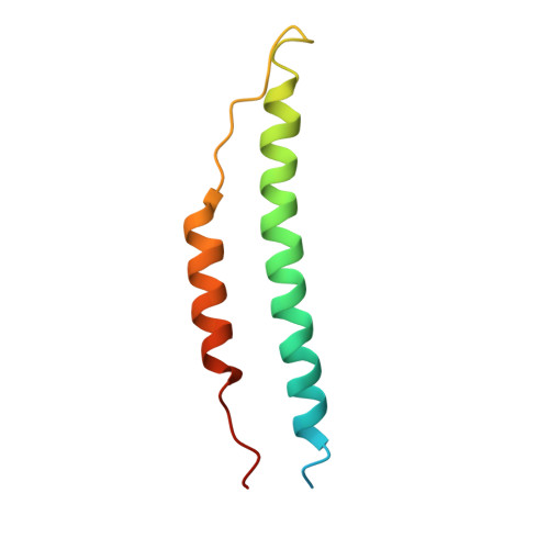
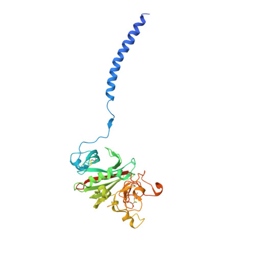
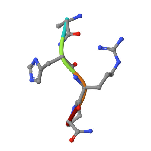
    The beta-chain amino-terminal sequences of all known mammalian fibrins begin with the sequence Gly-His-Arg-Pro- (GHRP-), but the homologous sequence in chicken fibrin begins with the sequence Ala-His-Arg-Pro- (AHRP-). Nonetheless, chicken fibrinogen binds the synthetic peptide GHRPam, and a previously reported crystal structure has revealed that the binding is in exact conformance with that observed for the human GHRPam-fragment D complex. We now report that human fibrinogen, which is known not to bind APRP, binds the synthetic peptide AHRPam. Moreover, a crystal structure of AHRPam complexed with fragment D from human fibrinogen shows that AHRPam binds exclusively to the beta-chain hole and, unlike GHRPam, not at all to the homologous gamma-chain hole. The difference can be attributed to the methyl group of the alanine residue clashing with a critical carboxyl group in the gammaC hole but being accommodated in the roomier betaC hole where the equivalent carboxyl is situated more flexibly.


  • Organizational Affiliation
    • Department of Chemistry and Biochemistry and Division of Biology, University of California at San Diego, La Jolla, California 92093-0314, USA. rdoolittle@ucsd.edu

Macromolecule Content 

  • Total Structure Weight: 171.05 kDa 
  • Atom Count: 10,770 
  • Modeled Residue Count: 1,334 
  • Deposited Residue Count: 1,486 
  • Unique protein chains: 4

Macromolecules

Find similar proteins by:|  3D Structure
Entity ID: 1
MoleculeChains  Sequence LengthOrganismDetailsImage
Fibrinogen alpha chain
A, D
87Homo sapiensMutation(s): 0 
UniProt & NIH Common Fund Data Resources
Find proteins for P02671 (Homo sapiens)
Explore P02671 
Go to UniProtKB:  P02671
PHAROS:  P02671
GTEx:  ENSG00000171560 
Entity Groups
Sequence Clusters30% Identity50% Identity70% Identity90% Identity95% Identity100% Identity
UniProt GroupP02671
Sequence Annotations
Expand
Reference Sequence
Find similar proteins by:|  3D Structure
Entity ID: 2
MoleculeChains  Sequence LengthOrganismDetailsImage
Fibrinogen beta chain
B, E
328Homo sapiensMutation(s): 0 
UniProt & NIH Common Fund Data Resources
Find proteins for P02675 (Homo sapiens)
Explore P02675 
Go to UniProtKB:  P02675
PHAROS:  P02675
GTEx:  ENSG00000171564 
Entity Groups
Sequence Clusters30% Identity50% Identity70% Identity90% Identity95% Identity100% Identity
UniProt GroupP02675
Glycosylation
Glycosylation Sites: 1Go to GlyGen: P02675-1
Sequence Annotations
Expand
Reference Sequence
Find similar proteins by:|  3D Structure
Entity ID: 3
MoleculeChains  Sequence LengthOrganismDetailsImage
Fibrinogen gamma chain
C, F
323Homo sapiensMutation(s): 0 
UniProt & NIH Common Fund Data Resources
Find proteins for P02679 (Homo sapiens)
Explore P02679 
Go to UniProtKB:  P02679
PHAROS:  P02679
GTEx:  ENSG00000171557 
Entity Groups
Sequence Clusters30% Identity50% Identity70% Identity90% Identity95% Identity100% Identity
UniProt GroupP02679
Sequence Annotations
Expand
Reference Sequence
Find similar proteins by:  Sequence   |   3D Structure  
Entity ID: 4
MoleculeChains  Sequence LengthOrganismDetailsImage
GLY-HIS-ARG-PRO-AMIDE peptide ligandG [auth I],
H [auth J]
5N/AMutation(s): 0 

Oligosaccharides

Help  
Entity ID: 5
MoleculeChains Length2D Diagram GlycosylationD Interactions
2-acetamido-2-deoxy-beta-D-glucopyranose-(1-4)-2-acetamido-2-deoxy-beta-D-glucopyranoseI [auth G]2N-Glycosylation
Glycosylation Resources
GlyTouCan: G42666HT
GlyCosmos: G42666HT
GlyGen: G42666HT
Entity ID: 6
MoleculeChains Length2D Diagram GlycosylationD Interactions
2-acetamido-2-deoxy-alpha-D-glucopyranose-(1-4)-2-acetamido-2-deoxy-beta-D-glucopyranoseJ [auth H]2N-Glycosylation
Glycosylation Resources
GlyTouCan: G07375KG
GlyCosmos: G07375KG
GlyGen: G07375KG

Experimental Data & Validation

Experimental Data

  • Method: X-RAY DIFFRACTION
  • Resolution: 2.70 Å
  • R-Value Free:  0.307 (Depositor), 0.305 (DCC) 
  • R-Value Work:  0.251 (Depositor), 0.243 (DCC) 
  • R-Value Observed: 0.244 (Depositor) 
Space Group: P 1 21 1
Unit Cell:
Length ( Å )Angle ( ˚ )
a = 105α = 90
b = 47.9β = 105.41
c = 171.4γ = 90
Software Package:
Software NamePurpose
AMoREphasing
CNSrefinement
ADSCdata collection
HKL-2000data scaling

Structure Validation

View Full Validation Report


Entry History 

Deposition Data

Revision History  (Full details and data files)

  • Version 1.0: 2006-12-05
    Type: Initial release
  • Version 1.1: 2008-05-01
    Changes: Version format compliance
  • Version 1.2: 2011-07-13
    Changes: Non-polymer description, Version format compliance
  • Version 2.0: 2020-07-29
    Type: Remediation
    Reason: Carbohydrate remediation
    Changes: Atomic model, Data collection, Database references, Derived calculations, Structure summary
  • Version 2.1: 2024-10-30
    Changes: Data collection, Database references, Structure summary