2H3G | pdb_00002h3g

Structure of the Type III Pantothenate Kinase (CoaX) from Bacillus Anthracis


Experimental Data Snapshot

  • Method: X-RAY DIFFRACTION
  • Resolution: 2.00 Å
  • R-Value Free: 
    0.258 (Depositor), 0.247 (DCC) 
  • R-Value Work: 
    0.215 (Depositor), 0.211 (DCC) 
  • R-Value Observed: 
    0.217 (Depositor) 

wwPDB Validation 3D Report Full Report

Validation slider image for 2H3G

This is version 1.4 of the entry. See complete history

Literature

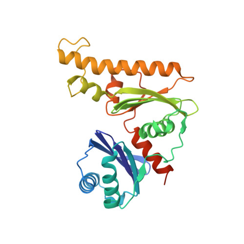
Structure of the Type III Pantothenate Kinase from Bacillus anthracis at 2.0 A Resolution: Implications for Coenzyme A-Dependent Redox Biology.

Nicely, N.I.Parsonage, D.Paige, C.Newton, G.L.Fahey, R.C.Leonardi, R.Jackowski, S.Mallett, T.C.Claiborne, A.

(2007) Biochemistry 46: 3234-3245

  • DOI: https://doi.org/10.1021/bi062299p
  • Primary Citation Related Structures: 
    2H3G

  • PubMed Abstract: 

    Coenzyme A (CoASH) is the major low-molecular weight thiol in Staphylococcus aureus and a number of other bacteria; the crystal structure of the S. aureus coenzyme A-disulfide reductase (CoADR), which maintains the reduced intracellular state of CoASH, has recently been reported [Mallett, T.C., Wallen, J.R., Karplus, P.A., Sakai, H., Tsukihara, T., and Claiborne, A. (2006) Biochemistry 45, 11278-89]. In this report we demonstrate that CoASH is the major thiol in Bacillus anthracis; a bioinformatics analysis indicates that three of the four proteins responsible for the conversion of pantothenate (Pan) to CoASH in Escherichia coli are conserved in B. anthracis. In contrast, a novel type III pantothenate kinase (PanK) catalyzes the first committed step in the biosynthetic pathway in B. anthracis; unlike the E. coli type I PanK, this enzyme is not subject to feedback inhibition by CoASH. The crystal structure of B. anthracis PanK (BaPanK), solved using multiwavelength anomalous dispersion data and refined at a resolution of 2.0 A, demonstrates that BaPanK is a new member of the Acetate and Sugar Kinase/Hsc70/Actin (ASKHA) superfamily. The Pan and ATP substrates have been modeled into the active-site cleft; in addition to providing a clear rationale for the absence of CoASH inhibition, analysis of the Pan-binding pocket has led to the development of two new structure-based motifs (the PAN and INTERFACE motifs). Our analyses also suggest that the type III PanK in the spore-forming B. anthracis plays an essential role in the novel thiol/disulfide redox biology of this category A biodefense pathogen.


  • Organizational Affiliation
    • Center for Structural Biology, Wake Forest University School of Medicine, Winston-Salem, North Carolina 27157, USA.

Macromolecule Content 

  • Total Structure Weight: 30.02 kDa 
  • Atom Count: 2,121 
  • Modeled Residue Count: 247 
  • Deposited Residue Count: 268 
  • Unique protein chains: 1

Macromolecules

Find similar proteins by:|  3D Structure
Entity ID: 1
MoleculeChains  Sequence LengthOrganismDetailsImage
BIOSYNTHETIC PROTEINA [auth X]268Bacillus anthracis str. AmesMutation(s): 0 
EC: 2.7.1.33
UniProt
Find proteins for Q81VX4 (Bacillus anthracis)
Explore Q81VX4 
Go to UniProtKB:  Q81VX4
Entity Groups
Sequence Clusters30% Identity50% Identity70% Identity90% Identity95% Identity100% Identity
UniProt GroupQ81VX4
Sequence Annotations
Expand
Reference Sequence

Small Molecules

Ligands 1 Unique
IDChains Name / Formula / InChI Key2D Diagram3D Interactions
EDO

Query on EDO


Download:Ideal Coordinates CCD File
B [auth X]1,2-ETHANEDIOL
C2 H6 O2
LYCAIKOWRPUZTN-UHFFFAOYSA-N

Experimental Data & Validation

Experimental Data

  • Method: X-RAY DIFFRACTION
  • Resolution: 2.00 Å
  • R-Value Free:  0.258 (Depositor), 0.247 (DCC) 
  • R-Value Work:  0.215 (Depositor), 0.211 (DCC) 
  • R-Value Observed: 0.217 (Depositor) 
Space Group: P 21 21 2
Unit Cell:
Length ( Å )Angle ( ˚ )
a = 67.852α = 90
b = 133.396β = 90
c = 41.138γ = 90
Software Package:
Software NamePurpose
DENZOdata reduction
SCALEPACKdata scaling
RESOLVEphasing
REFMACrefinement
PDB_EXTRACTdata extraction
CBASSdata collection
d*TREKdata scaling
SOLVEphasing

Structure Validation

View Full Validation Report


Entry History 

Deposition Data

  • Released Date: 2007-03-20 
  • Deposition Author(s): Nicely, N.I.

Revision History  (Full details and data files)

  • Version 1.0: 2007-03-20
    Type: Initial release
  • Version 1.1: 2008-04-01
    Changes: Version format compliance
  • Version 1.2: 2011-07-13
    Changes: Derived calculations, Source and taxonomy, Version format compliance
  • Version 1.3: 2017-10-18
    Changes: Refinement description
  • Version 1.4: 2024-02-14
    Changes: Data collection, Database references, Derived calculations