2GSM | pdb_00002gsm

Catalytic Core (Subunits I and II) of Cytochrome c oxidase from Rhodobacter sphaeroides


Experimental Data Snapshot

  • Method: X-RAY DIFFRACTION
  • Resolution: 2.00 Å
  • R-Value Free: 
    0.232 (Depositor), 0.234 (DCC) 
  • R-Value Work: 
    0.214 (Depositor), 0.215 (DCC) 
  • R-Value Observed: 
    0.214 (Depositor) 

wwPDB Validation 3D Report Full Report

Validation slider image for 2GSM

Ligand Structure Quality Assessment 


This is version 1.4 of the entry. See complete history

Literature

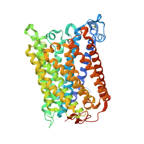
Identification of conserved lipid/detergent-binding sites in a high-resolution structure of the membrane protein cytochrome c oxidase.

Qin, L.Hiser, C.Mulichak, A.Garavito, R.M.Ferguson-Miller, S.

(2006) Proc Natl Acad Sci U S A 103: 16117-16122

  • DOI: https://doi.org/10.1073/pnas.0606149103
  • Primary Citation Related Structures: 
    2GSM

  • PubMed Abstract: 

    Well ordered reproducible crystals of cytochrome c oxidase (CcO) from Rhodobacter sphaeroides yield a previously unreported structure at 2.0 A resolution that contains the two catalytic subunits and a number of alkyl chains of lipids and detergents. Comparison with crystal structures of other bacterial and mammalian CcOs reveals that the positions occupied by native membrane lipids and detergent substitutes are highly conserved, along with amino acid residues in their vicinity, suggesting a more prevalent and specific role of lipid in membrane protein structure than often envisioned. Well defined detergent head groups (maltose) are found associated with aromatic residues in a manner similar to phospholipid head groups, likely contributing to the success of alkyl glycoside detergents in supporting membrane protein activity and crystallizability. Other significant features of this structure include the following: finding of a previously unreported crystal contact mediated by cadmium and an engineered histidine tag; documentation of the unique His-Tyr covalent linkage close to the active site; remarkable conservation of a chain of waters in one proton pathway (D-path); and discovery of an inhibitory cadmium-binding site at the entrance to another proton path (K-path). These observations provide important insight into CcO structure and mechanism, as well as the significance of bound lipid in membrane proteins.


  • Organizational Affiliation
    • Department of Biochemistry and Molecular Biology, Michigan State University, East Lansing, MI 48824, USA.

Macromolecule Content 

  • Total Structure Weight: 196.56 kDa 
  • Atom Count: 13,644 
  • Modeled Residue Count: 1,581 
  • Deposited Residue Count: 1,656 
  • Unique protein chains: 2

Macromolecules

Find similar proteins by:|  3D Structure
Entity ID: 1
MoleculeChains  Sequence LengthOrganismDetailsImage
Cytochrome c oxidase subunit 1
A, C
566Cereibacter sphaeroidesMutation(s): 0 
Gene Names: ctaD
EC: 1.9.3.1 (PDB Primary Data), 7.1.1.9 (UniProt)
Membrane Entity: Yes 
UniProt
Find proteins for P33517 (Cereibacter sphaeroides)
Explore P33517 
Go to UniProtKB:  P33517
Entity Groups
Sequence Clusters30% Identity50% Identity70% Identity90% Identity95% Identity100% Identity
UniProt GroupP33517
Sequence Annotations
Expand
Reference Sequence
Find similar proteins by:|  3D Structure
Entity ID: 2
MoleculeChains  Sequence LengthOrganismDetailsImage
Cytochrome c oxidase subunit 2
B, D
262Cereibacter sphaeroidesMutation(s): 0 
Gene Names: ctaCcoxIIctaB
EC: 1.9.3.1 (PDB Primary Data), 7.1.1.9 (UniProt)
Membrane Entity: Yes 
UniProt
Find proteins for Q03736 (Cereibacter sphaeroides)
Explore Q03736 
Go to UniProtKB:  Q03736
Entity Groups
Sequence Clusters30% Identity50% Identity70% Identity90% Identity95% Identity100% Identity
UniProt GroupQ03736
Sequence Annotations
Expand
Reference Sequence

Small Molecules

Ligands 8 Unique
IDChains Name / Formula / InChI Key2D Diagram3D Interactions
HEA

Query on HEA


Download:Ideal Coordinates CCD File
GA [auth C],
HA [auth C],
L [auth A],
M [auth A]
HEME-A
C49 H56 Fe N4 O6
ZGGYGTCPXNDTRV-PRYGPKJJSA-L
DMU

Query on DMU


Download:Ideal Coordinates CCD File
AA [auth C]
BA [auth C]
E [auth A]
F [auth A]
G [auth A]
AA [auth C],
BA [auth C],
E [auth A],
F [auth A],
G [auth A],
MA [auth D],
NA [auth D],
S [auth B],
T [auth B],
Z [auth C]
DECYL-BETA-D-MALTOPYRANOSIDE
C22 H42 O11
WOQQAWHSKSSAGF-WXFJLFHKSA-N
TRD

Query on TRD


Download:Ideal Coordinates CCD File
IA [auth C]
JA [auth C]
KA [auth C]
LA [auth C]
N [auth A]
IA [auth C],
JA [auth C],
KA [auth C],
LA [auth C],
N [auth A],
O [auth A],
P [auth A],
Q [auth A],
R [auth A],
SA [auth D],
TA [auth D],
Y [auth B]
TRIDECANE
C13 H28
IIYFAKIEWZDVMP-UHFFFAOYSA-N
CD

Query on CD


Download:Ideal Coordinates CCD File
QA [auth D],
RA [auth D],
W [auth B],
X [auth B]
CADMIUM ION
Cd
WLZRMCYVCSSEQC-UHFFFAOYSA-N
CU

Query on CU


Download:Ideal Coordinates CCD File
CA [auth C]
H [auth A]
OA [auth D]
PA [auth D]
U [auth B]
CA [auth C],
H [auth A],
OA [auth D],
PA [auth D],
U [auth B],
V [auth B]
COPPER (II) ION
Cu
JPVYNHNXODAKFH-UHFFFAOYSA-N
CA

Query on CA


Download:Ideal Coordinates CCD File
EA [auth C],
J [auth A]
CALCIUM ION
Ca
BHPQYMZQTOCNFJ-UHFFFAOYSA-N
MG

Query on MG


Download:Ideal Coordinates CCD File
DA [auth C],
I [auth A]
MAGNESIUM ION
Mg
JLVVSXFLKOJNIY-UHFFFAOYSA-N
OH

Query on OH


Download:Ideal Coordinates CCD File
FA [auth C],
K [auth A]
HYDROXIDE ION
H O
XLYOFNOQVPJJNP-UHFFFAOYSA-M

Experimental Data & Validation

Experimental Data

  • Method: X-RAY DIFFRACTION
  • Resolution: 2.00 Å
  • R-Value Free:  0.232 (Depositor), 0.234 (DCC) 
  • R-Value Work:  0.214 (Depositor), 0.215 (DCC) 
  • R-Value Observed: 0.214 (Depositor) 
Space Group: P 21 21 21
Unit Cell:
Length ( Å )Angle ( ˚ )
a = 125.02α = 90
b = 131.639β = 90
c = 176.802γ = 90
Software Package:
Software NamePurpose
REFMACrefinement
MAR345data collection
XDSdata scaling
PHASERphasing

Structure Validation

View Full Validation Report


Ligand Structure Quality Assessment 


Entry History 

Deposition Data

Revision History  (Full details and data files)

  • Version 1.0: 2006-10-10
    Type: Initial release
  • Version 1.1: 2007-10-16
    Changes: Version format compliance
  • Version 1.2: 2011-07-13
    Changes: Derived calculations, Version format compliance
  • Version 1.3: 2017-10-18
    Changes: Advisory, Refinement description
  • Version 1.4: 2024-11-13
    Changes: Advisory, Data collection, Database references, Derived calculations, Structure summary