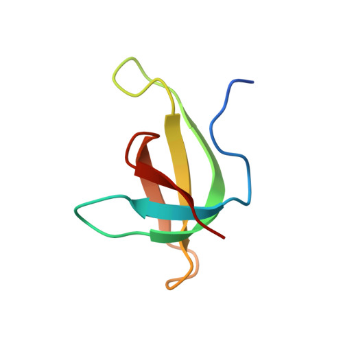
High-resolution structure of a plasmid-encoded dihydrofolate reductase: pentagonal network of water molecules in the D(2)-symmetric active site.
Narayana, N.(2006) Acta Crystallogr D Biol Crystallogr 62: 695-706
- PubMed: 16790925 Search on PubMed
- DOI: https://doi.org/10.1107/S0907444906014764
- Primary Citation Related Structures:
2GQV - PubMed Abstract:
R67 plasmid-encoded dihydrofolate reductase (R67 DHFR) is an NADPH-dependent homotetrameric enzyme that catalyzes the reduction of dihydrofolate to tetrahydrofolate. The amino-acid sequence and molecular architecture of R67 DHFR and its inhibitory properties toward folate analogues are different from those of chromosomal DHFR. Here, the crystal structure of R67 DHFR refined using 1.1 A resolution data is presented. Blocked full-matrix least-squares refinement without restraints resulted in a final R factor of 11.4%. The anisotropic atomic displacement parameters analyzed by Rosenfield matrices and translation-libration-screw validation suggested four quasi-rigid domains. A total of ten Calpha-H...O hydrogen bonds were identified between the beta-strands. There is reasonable structural evidence that His62 is not protonated in the tetramer, which is in accord with previous pH-profile studies. The side chain of Gln67 that protrudes into the active site exhibits dual conformation, a feature noticed for the first time owing to the availability of atomic resolution data. The R67 DHFR active site is unique: it has D2 symmetry and is a large active site with a pentagonal network of water molecules and exposure of backbone atoms to solvent; the central pore is favorable for planar ring-stacking interactions. The geometrical shape, overall symmetry, local asymmetry and waters appear to dominate the binding of ligands, catalysis and inhibition.
- Department of Biochemistry, Case Western Reserve University, Cleveland, OH 44106, USA. nxn17@case.edu
Organizational Affiliation: