Systematic structural studies of iron superoxide dismutases from human parasites and a statistical coupling analysis of metal binding specificity
Bachega, J.F., Navarro, M.V., Bleicher, L., Bortoleto-Bugs, R.K., Dive, D., Hoffmann, P., Viscogliosi, E., Garratt, R.C.(2009) Proteins 77: 26-37
- PubMed: 19384994 Search on PubMed
- DOI: https://doi.org/10.1002/prot.22412
- Primary Citation Related Structures:
2GOJ, 2GPC, 3ESF - PubMed Abstract:
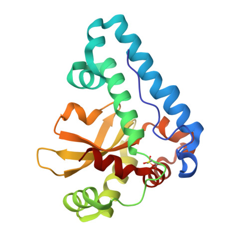
Superoxide dismutases (SODs) are a crucial class of enzymes in the combat against intracellular free radical damage. They eliminate superoxide radicals by converting them into hydrogen peroxide and oxygen. In spite of their very different life cycles and infection strategies, the human parasites Plasmodium falciparum, Trypanosoma cruzi and Trypanosoma brucei are known to be sensitive to oxidative stress. Thus the parasite Fe-SODs have become attractive targets for novel drug development. Here we report the crystal structures of FeSODs from the trypanosomes T. brucei at 2.0 A and T. cruzi at 1.9 A resolution, and that from P. falciparum at a higher resolution (2.0 A) to that previously reported. The homodimeric enzymes are compared to the related human MnSOD with particular attention to structural aspects which are relevant for drug design. Although the structures possess a very similar overall fold, differences between the enzymes at the entrance to the channel which leads to the active site could be identified. These lead to a slightly broader and more positively charged cavity in the parasite enzymes. Furthermore, a statistical coupling analysis (SCA) for the whole Fe/MnSOD family reveals different patterns of residue coupling for Mn and Fe SODs, as well as for the dimeric and tetrameric states. In both cases, the statistically coupled residues lie adjacent to the conserved core surrounding the metal center and may be expected to be responsible for its fine tuning, leading to metal ion specificity.
- Institute of Physics of São Carlos, University of São Paulo, CEP 13560-970, São Carlos, SP, Brazil.
Organizational Affiliation: