2GM4 | pdb_00002gm4

An activated, tetrameric gamma-delta resolvase: Hin chimaera bound to cleaved DNA


Experimental Data Snapshot

  • Method: X-RAY DIFFRACTION
  • Resolution: 3.50 Å
  • R-Value Free: 
    0.323 (Depositor), 0.323 (DCC) 
  • R-Value Work: 
    0.280 (Depositor), 0.277 (DCC) 
  • R-Value Observed: 
    0.282 (Depositor) 

Starting Model: experimental
View more details

wwPDB Validation 3D Report Full Report

Validation slider image for 2GM4

This is version 1.5 of the entry. See complete history

Literature

Implications of structures of synaptic tetramers of gamma delta resolvase for the mechanism of recombination.

Kamtekar, S.Ho, R.S.Cocco, M.J.Li, W.Wenwieser, S.V.Boocock, M.R.Grindley, N.D.Steitz, T.A.

(2006) Proc Natl Acad Sci U S A 103: 10642-10647

  • DOI: https://doi.org/10.1073/pnas.0604062103
  • Primary Citation Related Structures: 
    2GM4, 2GM5

  • PubMed Abstract: 

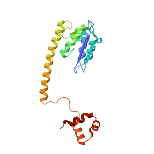
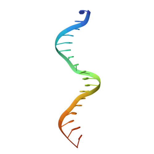
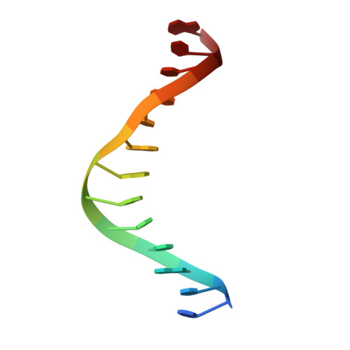
    The structures of two mutants of the site-specific recombinase, gammadelta resolvase, that form activated tetramers have been determined. One, at 3.5-A resolution, forms a synaptic intermediate of resolvase that is covalently linked to two cleaved DNAs, whereas the other is of an unliganded structure determined at 2.1-A resolution. Comparisons of the four known tetrameric resolvase structures show that the subunits interact through the formation of a common core of four helices. The N-terminal halves of these helices superimpose well on each other, whereas the orientations of their C termini are more variable. The catalytic domains of resolvase in the unliganded structure are arranged asymmetrically, demonstrating that their positions can move substantially while preserving the four-helix core that forms the tetramer. These results suggest that the precleavage synaptic tetramer of gammadelta resolvase, whose structure is not known, may be formed by a similar four-helix core, but differ in the relative orientations of its catalytic and DNA-binding domains.


  • Organizational Affiliation
    • Departments of Molecular Biophysics, and Howard Hughes Medical Institute, Yale University, New Haven, CT 06520-8114, USA.

Macromolecule Content 

  • Total Structure Weight: 74.39 kDa 
  • Atom Count: 4,132 
  • Modeled Residue Count: 423 
  • Deposited Residue Count: 476 
  • Unique protein chains: 1
  • Unique nucleic acid chains: 2

Macromolecules


Find similar proteins by:|  3D Structure
Entity ID: 3
MoleculeChains  Sequence LengthOrganismDetailsImage
Transposon gamma-delta resolvaseG [auth A],
H [auth B]
183Escherichia coliMutation(s): 9 
Gene Names: tnpR
UniProt
Find proteins for P03012 (Escherichia coli (strain K12))
Explore P03012 
Go to UniProtKB:  P03012
Entity Groups
Sequence Clusters30% Identity50% Identity70% Identity90% Identity95% Identity100% Identity
UniProt GroupP03012
Sequence Annotations
Expand
Reference Sequence
Find similar nucleic acids by:  Sequence
Entity ID: 1
MoleculeChains LengthOrganismImage
5'-D(*CP*AP*GP*TP*GP*TP*CP*CP*GP*AP*TP*AP*AP*TP*TP*TP*AP*TP*AP*AP*A)-3'A [auth X],
B [auth Z],
D [auth J],
E [auth I]
21N/A
Sequence Annotations
Expand
Reference Sequence
Find similar nucleic acids by:  Sequence
Entity ID: 2
MoleculeChains LengthOrganismImage
5'-D(*TP*TP*AP*TP*CP*GP*GP*AP*CP*AP*CP*TP*G)-3'C [auth Y],
F [auth K]
13N/A
Sequence Annotations
Expand
Reference Sequence

Experimental Data & Validation

Experimental Data

  • Method: X-RAY DIFFRACTION
  • Resolution: 3.50 Å
  • R-Value Free:  0.323 (Depositor), 0.323 (DCC) 
  • R-Value Work:  0.280 (Depositor), 0.277 (DCC) 
  • R-Value Observed: 0.282 (Depositor) 
Space Group: P 21 21 2
Unit Cell:
Length ( Å )Angle ( ˚ )
a = 110.138α = 90
b = 137.687β = 90
c = 83.473γ = 90
Software Package:
Software NamePurpose
REFMACrefinement
ADSCdata collection
SCALEPACKdata scaling
CNSphasing

Structure Validation

View Full Validation Report


Entry History 

Deposition Data

Revision History  (Full details and data files)

  • Version 1.0: 2006-06-27
    Type: Initial release
  • Version 1.1: 2008-05-01
    Changes: Version format compliance
  • Version 1.2: 2011-07-13
    Changes: Advisory, Version format compliance
  • Version 1.3: 2021-10-20
    Changes: Database references, Derived calculations
  • Version 1.4: 2023-08-30
    Changes: Data collection, Refinement description
  • Version 1.5: 2024-11-20
    Changes: Structure summary