Structural Insights into the Non-additivity Effects in the Sequence-to-Reactivity Algorithm for Serine Peptidases and their Inhibitors.
Lee, T.W., Qasim, M.A., Laskowski, M., James, M.N.(2007) J Mol Biology 367: 527-546
- PubMed: 17266986 Search on PubMed
- DOI: https://doi.org/10.1016/j.jmb.2007.01.008
- Primary Citation Related Structures:
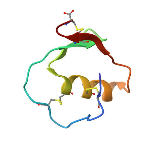
2GKR, 2GKT, 2GKV - PubMed Abstract:
Sequence-to-reactivity algorithms (SRAs) for proteins have the potential of being broadly applied in molecular design. Recently, Laskowski et al. have reported an additivity-based SRA that accurately predicts most of the standard free energy changes of association for variants of turkey ovomucoid third domain (OMTKY3) with six serine peptidases, one of which is streptogrisin B (commonly known as Streptomyces griseus peptidase B, SGPB). Non-additivity effects for residues 18I and 32I, and for residues 20I and 32I of OMTKY3 occurred when the associations with SGPB were predicted using the SRA. To elucidate precisely the mechanics of these non-additivity effects in structural terms, we have determined the crystal structures of the unbound OMTKY3 (with Gly32I as in the wild-type amino acid sequence) at a resolution of 1.16 A, the unbound Ala32I variant of OMTKY3 at a resolution of 1.23 A, and the SGPB:OMTKY3-Ala32I complex (equilibrium association constant K(a)=7.1x10(9) M(-1) at 21(+/-2) C degrees, pH 8.3) at a resolution of 1.70 A. Extensive comparisons with the crystal structure of the unbound OMTKY3 confirm our understanding of some previously addressed non-additivity effects. Unexpectedly, SGPB and OMTKY3-Ala32I form a 1:2 complex in the crystal. Comparison with the SGPB:OMTKY3 complex shows a conformational change in the SGPB:OMTKY3-Ala32I complex, resulting from a hinged rigid-body rotation of the inhibitor caused by the steric hindrance between the methyl group of Ala32IA of the inhibitor and Pro192BE of the peptidase. This perturbs the interactions among residues 18I, 20I, 32I and 36I of the inhibitor, probably resulting in the above non-additivity effects. This conformational change also introduces residue 10I as an additional hyper-variable contact residue to the SRA.
- Group in Protein Structure and Function, Department of Biochemistry, University of Alberta, Edmonton, Alberta, Canada T6G 2H7.
Organizational Affiliation: