2GEE | pdb_00002gee

Crystal Structure of Human Type III Fibronectin Extradomain B and Domain 8


Experimental Data Snapshot

  • Method: X-RAY DIFFRACTION
  • Resolution: 2.01 Å
  • R-Value Free: 
    0.301 (Depositor) 
  • R-Value Work: 
    0.249 (Depositor) 
  • R-Value Observed: 
    0.249 (Depositor) 

Starting Model: experimental
View more details

wwPDB Validation 3D Report Full Report

Validation slider image for 2GEE

This is version 1.3 of the entry. See complete history

Literature

Structural insights into fibronectin type III domain-mediated signaling.

Bencharit, S.Cui, C.B.Siddiqui, A.Howard-Williams, E.L.Sondek, J.Zuobi-Hasona, K.Aukhil, I.

(2007) J Mol Biology 367: 303-309

  • DOI: https://doi.org/10.1016/j.jmb.2006.10.017
  • Primary Citation Related Structures: 
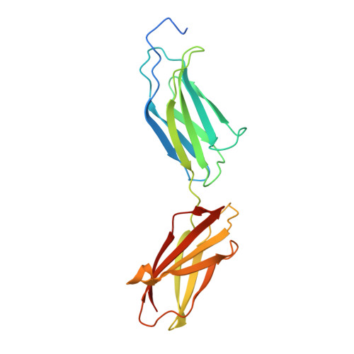
    2GEE

  • PubMed Abstract: 

    The alternatively spliced type III extradomain B (EIIIB) of fibronectin (FN) is expressed only during embryogenesis, wound healing and tumorigenesis. The biological function of this domain is unclear. We describe here the first crystal structure of the interface between alternatively spliced EIIIB and its adjacent FN type III domain 8 (FN B-8). The opened CC' loop of EIIIB, and the rotation and tilt of EIIIB allow good access to the FG loop of FN-8, which is normally hindered by the CC' loop of FN-7. In addition, the AGEGIP sequence of the CC'' loop of EIIIB replaces the NGQQGN sequence of the CC' loop of FN-7. Finally, the CC'' loop of EIIIB forms an acidic groove with FN-8. These structural findings warrant future studies directed at identifying potential binding partners for FN B-8 interface, linking EIIIB to skeletal and cartilaginous development, wound healing, and tumorigenesis, respectively.


  • Organizational Affiliation
    • Department of Prosthodontics, School of Dentistry, University of North Carolina, Chapel Hill, NC 27599, USA.

Macromolecule Content 

  • Total Structure Weight: 22.1 kDa 
  • Atom Count: 1,827 
  • Modeled Residue Count: 188 
  • Deposited Residue Count: 203 
  • Unique protein chains: 1

Macromolecules

Find similar proteins by:|  3D Structure
Entity ID: 1
MoleculeChains  Sequence LengthOrganismDetailsImage
hypothetical protein203Homo sapiensMutation(s): 0 
Gene Names: DKFZp686O1166
UniProt & NIH Common Fund Data Resources
Find proteins for P02751 (Homo sapiens)
Explore P02751 
Go to UniProtKB:  P02751
PHAROS:  P02751
GTEx:  ENSG00000115414 
Entity Groups
Sequence Clusters30% Identity50% Identity70% Identity90% Identity95% Identity100% Identity
UniProt GroupP02751
Sequence Annotations
Expand
Reference Sequence

Experimental Data & Validation

Experimental Data

  • Method: X-RAY DIFFRACTION
  • Resolution: 2.01 Å
  • R-Value Free:  0.301 (Depositor) 
  • R-Value Work:  0.249 (Depositor) 
  • R-Value Observed: 0.249 (Depositor) 
Space Group: P 21 21 21
Unit Cell:
Length ( Å )Angle ( ˚ )
a = 43.407α = 90
b = 55.288β = 90
c = 74.903γ = 90
Software Package:
Software NamePurpose
CNSrefinement
CrystalCleardata reduction
HKL-2000data scaling
AMoREphasing

Structure Validation

View Full Validation Report


Entry History 

Deposition Data

Revision History  (Full details and data files)

  • Version 1.0: 2006-10-17
    Type: Initial release
  • Version 1.1: 2008-05-01
    Changes: Version format compliance
  • Version 1.2: 2011-07-13
    Changes: Version format compliance
  • Version 1.3: 2023-08-30
    Changes: Data collection, Database references, Refinement description