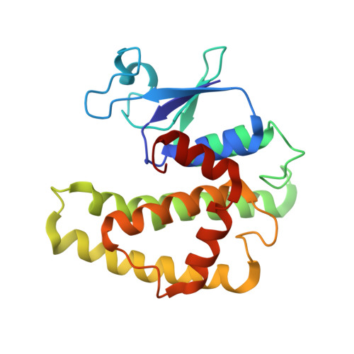
2GDR

Crystal structure of a bacterial glutathione transferase


Experimental Data Snapshot

  • Method: X-RAY DIFFRACTION
  • Resolution: 2.10 Å
  • R-Value Free: 0.204 
  • R-Value Work: 0.161 
  • R-Value Observed: 0.161 

Starting Model: experimental
View more details

wwPDB Validation   3D Report Full Report


Ligand Structure Quality Assessment 


This is version 1.4 of the entry. See complete history


Literature

Structures of Ternary Complexes of BphK, a Bacterial Glutathione S-Transferase That Reductively Dechlorinates Polychlorinated Biphenyl Metabolites.

Tocheva, E.I.Fortin, P.D.Eltis, L.D.Murphy, M.E.

(2006) J Biol Chem 281: 30933-30940

  • DOI: https://doi.org/10.1074/jbc.M603125200
  • Primary Citation of Related Structures:  
    2DSA, 2GDR

  • PubMed Abstract: 

    Prokaryotic glutathione S-transferases are as diverse as their eukaryotic counterparts but are much less well characterized. BphK from Burkholderia xenovorans LB400 consumes two GSH molecules to reductively dehalogenate chlorinated 2-hydroxy-6-oxo-6-phenyl-2,4-dienoates (HOPDAs), inhibitory polychlorinated biphenyl metabolites. Crystallographic structures of two ternary complexes of BphK were solved to a resolution of 2.1A. In the BphK-GSH-HOPDA complex, GSH and HOPDA molecules occupy the G- and H-subsites, respectively. The thiol nucleophile of the GSH molecule is positioned for SN2 attack at carbon 3 of the bound HOPDA. The respective sulfur atoms of conserved Cys-10 and the bound GSH are within 3.0A, consistent with product release and the formation of a mixed disulfide intermediate. In the BphK-(GSH)2 complex, a GSH molecule occupies each of the two subsites. The three sulfur atoms of the two GSH molecules and Cys-10 are aligned suitably for a disulfide exchange reaction that would regenerate the resting enzyme and yield disulfide-linked GSH molecules. A second conserved residue, His-106, is adjacent to the thiols of Cys-10 and the GSH bound to the G-subsite and thus may stabilize a transition state in the disulfide exchange reaction. Overall, the structures support and elaborate a proposed dehalogenation mechanism for BphK and provide insight into the plasticity of the H-subsite.


  • Organizational Affiliation

    Department of Microbiology and Immunology, University of British Columbia, Vancouver, British Columbia V6T 1Z3, Canada.


Macromolecules
Find similar proteins by:  (by identity cutoff)  |  3D Structure
Entity ID: 1
MoleculeChains Sequence LengthOrganismDetailsImage
glutathione S-transferase
A, B, C, D, E
A, B, C, D, E, F
202Paraburkholderia xenovorans LB400Mutation(s): 0 
Gene Names: bphK
EC: 2.5.1.18
UniProt
Find proteins for Q59721 (Paraburkholderia xenovorans (strain LB400))
Explore Q59721 
Go to UniProtKB:  Q59721
Entity Groups  
Sequence Clusters30% Identity50% Identity70% Identity90% Identity95% Identity100% Identity
UniProt GroupQ59721
Sequence Annotations
Expand
  • Reference Sequence
Experimental Data & Validation

Experimental Data

  • Method: X-RAY DIFFRACTION
  • Resolution: 2.10 Å
  • R-Value Free: 0.204 
  • R-Value Work: 0.161 
  • R-Value Observed: 0.161 
  • Space Group: P 65
Unit Cell:
Length ( Å )Angle ( ˚ )
a = 103.44α = 90
b = 103.44β = 90
c = 262.28γ = 120
Software Package:
Software NamePurpose
DENZOdata reduction
SCALEPACKdata scaling
CNSrefinement
CNSphasing

Structure Validation

View Full Validation Report


Ligand Structure Quality Assessment 


Entry History 

Deposition Data

Revision History  (Full details and data files)

  • Version 1.0: 2006-08-22
    Type: Initial release
  • Version 1.1: 2008-05-01
    Changes: Version format compliance
  • Version 1.2: 2011-07-13
    Changes: Source and taxonomy, Version format compliance
  • Version 1.3: 2011-12-14
    Changes: Non-polymer description
  • Version 1.4: 2023-08-30
    Changes: Data collection, Database references, Derived calculations, Refinement description