2GCG | pdb_00002gcg

Ternary Crystal Structure of Human Glyoxylate Reductase/Hydroxypyruvate Reductase


Experimental Data Snapshot

  • Method: X-RAY DIFFRACTION
  • Resolution: 2.20 Å
  • R-Value Free: 
    0.282 (Depositor), 0.286 (DCC) 
  • R-Value Work: 
    0.201 (Depositor), 0.208 (DCC) 
  • R-Value Observed: 
    0.205 (Depositor) 

Starting Model: experimental
View more details

wwPDB Validation 3D Report Full Report

Validation slider image for 2GCG

Ligand Structure Quality Assessment 


This is version 1.4 of the entry. See complete history

Literature

Structural basis of substrate specificity in human glyoxylate reductase/hydroxypyruvate reductase

Booth, M.P.S.Conners, R.Rumsby, G.Brady, R.L.

(2006) J Mol Biology 360: 178-189

  • DOI: https://doi.org/10.1016/j.jmb.2006.05.018
  • Primary Citation Related Structures: 
    2GCG, 2WWR

  • PubMed Abstract: 

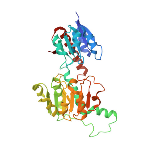
    Human glyoxylate reductase/hydroxypyruvate reductase (GRHPR) is a D-2-hydroxy-acid dehydrogenase that plays a critical role in the removal of the metabolic by-product glyoxylate from within the liver. Deficiency of this enzyme is the underlying cause of primary hyperoxaluria type 2 (PH2) and leads to increased urinary oxalate levels, formation of kidney stones and renal failure. Here we describe the crystal structure of human GRHPR at 2.2 A resolution. There are four copies of GRHPR in the crystallographic asymmetric unit: in each homodimer, one subunit forms a ternary (enzyme+NADPH+reduced substrate) complex, and the other a binary (enzyme+NADPH) form. The spatial arrangement of the two enzyme domains is the same in binary and ternary forms. This first crystal structure of a true ternary complex of an enzyme from this family demonstrates the relationship of substrate and catalytic residues within the active site, confirming earlier proposals of the mode of substrate binding, stereospecificity and likely catalytic mechanism for these enzymes. GRHPR has an unusual substrate specificity, preferring glyoxylate and hydroxypyruvate, but not pyruvate. A tryptophan residue (Trp141) from the neighbouring subunit of the dimer is projected into the active site region and appears to contribute to the selectivity for hydroxypyruvate. This first crystal structure of a human GRHPR enzyme also explains the deleterious effects of naturally occurring missense mutations of this enzyme that lead to PH2.


  • Organizational Affiliation
    • Department of Biochemistry, University of Bristol, Bristol BS8 1TD, UK.

Macromolecule Content 

  • Total Structure Weight: 147.15 kDa 
  • Atom Count: 10,701 
  • Modeled Residue Count: 1,294 
  • Deposited Residue Count: 1,320 
  • Unique protein chains: 1

Macromolecules

Find similar proteins by:|  3D Structure
Entity ID: 1
MoleculeChains  Sequence LengthOrganismDetailsImage
Glyoxylate reductase/hydroxypyruvate reductase
A, B, C, D
330Homo sapiensMutation(s): 0 
EC: 1.1.1.79 (PDB Primary Data), 1.1.1.81 (UniProt)
UniProt & NIH Common Fund Data Resources
Find proteins for Q9UBQ7 (Homo sapiens)
Explore Q9UBQ7 
Go to UniProtKB:  Q9UBQ7
PHAROS:  Q9UBQ7
GTEx:  ENSG00000137106 
Entity Groups
Sequence Clusters30% Identity50% Identity70% Identity90% Identity95% Identity100% Identity
UniProt GroupQ9UBQ7
Sequence Annotations
Expand
Reference Sequence

Small Molecules

Ligands 3 Unique
IDChains Name / Formula / InChI Key2D Diagram3D Interactions
NDP

Query on NDP


Download:Ideal Coordinates CCD File
E [auth A],
H [auth B],
J [auth C],
O [auth D]
NADPH DIHYDRO-NICOTINAMIDE-ADENINE-DINUCLEOTIDE PHOSPHATE
C21 H30 N7 O17 P3
ACFIXJIJDZMPPO-NNYOXOHSSA-N
DGY

Query on DGY


Download:Ideal Coordinates CCD File
F [auth A],
K [auth C]
(2R)-2,3-DIHYDROXYPROPANOIC ACID
C3 H6 O4
RBNPOMFGQQGHHO-UWTATZPHSA-N
SO4

Query on SO4


Download:Ideal Coordinates CCD File
G [auth B],
I [auth C],
L [auth D],
M [auth D],
N [auth D]
SULFATE ION
O4 S
QAOWNCQODCNURD-UHFFFAOYSA-L

Experimental Data & Validation

Experimental Data

  • Method: X-RAY DIFFRACTION
  • Resolution: 2.20 Å
  • R-Value Free:  0.282 (Depositor), 0.286 (DCC) 
  • R-Value Work:  0.201 (Depositor), 0.208 (DCC) 
  • R-Value Observed: 0.205 (Depositor) 
Space Group: P 1 21 1
Unit Cell:
Length ( Å )Angle ( ˚ )
a = 76.568α = 90
b = 66.877β = 98.22
c = 149.78γ = 90
Software Package:
Software NamePurpose
DENZOdata reduction
SCALEPACKdata scaling
REFMACrefinement
PDB_EXTRACTdata extraction
HKL-2000data reduction
PHASERphasing

Structure Validation

View Full Validation Report


Ligand Structure Quality Assessment 


Entry History 

Deposition Data

Revision History  (Full details and data files)

  • Version 1.0: 2006-07-18
    Type: Initial release
  • Version 1.1: 2008-05-01
    Changes: Version format compliance
  • Version 1.2: 2011-07-13
    Changes: Advisory, Version format compliance
  • Version 1.3: 2017-10-18
    Changes: Refinement description
  • Version 1.4: 2023-10-25
    Changes: Data collection, Database references, Derived calculations, Refinement description