2GBQ

SOLUTION NMR STRUCTURE OF THE GRB2 N-TERMINAL SH3 DOMAIN COMPLEXED WITH A TEN-RESIDUE PEPTIDE DERIVED FROM SOS DIRECT REFINEMENT AGAINST NOES, J-COUPLINGS, AND 1H AND 13C CHEMICAL SHIFTS, 15 STRUCTURES


Experimental Data Snapshot

  • Method: SOLUTION NMR
  • Conformers Calculated: 200 
  • Conformers Submitted: 15 

wwPDB Validation   3D Report Full Report


This is version 1.3 of the entry. See complete history


Literature

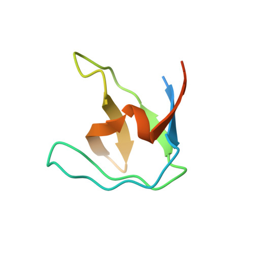
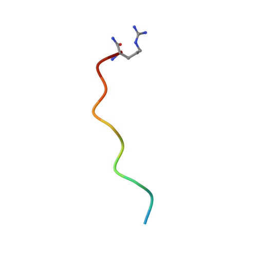
Solution structure of the Grb2 N-terminal SH3 domain complexed with a ten-residue peptide derived from SOS: direct refinement against NOEs, J-couplings and 1H and 13C chemical shifts.

Wittekind, M.Mapelli, C.Lee, V.Goldfarb, V.Friedrichs, M.S.Meyers, C.A.Mueller, L.

(1997) J Mol Biol 267: 933-952

  • DOI: https://doi.org/10.1006/jmbi.1996.0886
  • Primary Citation of Related Structures:  
    1GBQ, 2GBQ, 3GBQ, 4GBQ

  • PubMed Abstract: 
  • Refined ensembles of solution structures have been calculated for the N-terminal SH3 domain of Grb2 (N-SH3) complexed with the ac-VPPPVPPRRR-nh2 peptide derived from residues 1135 to 1144 of the mouse SOS-1 sequence. NMR spectra obtained from different combinations of both 13C-15N-labeled and unlabeled N-SH3 and SOS peptide fragment were used to obtain stereo-assignments for pro-chiral groups of the peptide, angle restraints via heteronuclear coupling constants, and complete 1H, 13C, and 15N resonance assignments for both molecules ...

    Refined ensembles of solution structures have been calculated for the N-terminal SH3 domain of Grb2 (N-SH3) complexed with the ac-VPPPVPPRRR-nh2 peptide derived from residues 1135 to 1144 of the mouse SOS-1 sequence. NMR spectra obtained from different combinations of both 13C-15N-labeled and unlabeled N-SH3 and SOS peptide fragment were used to obtain stereo-assignments for pro-chiral groups of the peptide, angle restraints via heteronuclear coupling constants, and complete 1H, 13C, and 15N resonance assignments for both molecules. One ensemble of structures was calculated using conventional methods while a second ensemble was generated by including additional direct refinements against both 1H and 13C(alpha)/13C(beta) chemical shifts. In both ensembles, the protein:peptide interface is highly resolved, reflecting the inclusion of 110 inter-molecular nuclear Overhauser enhancement (NOE) distance restraints. The first and second peptide-binding sub-sites of N-SH3 interact with structurally well-defined portions of the peptide. These interactions include hydrogen bonds and extensive hydrophobic contacts. In the third highly acidic sub-site, the conformation of the peptide Arg8 side-chain is partially ordered by a set of NOE restraints to the Trp36 ring protons. Overall, several lines of evidence point to dynamical averaging of peptide and N-SH3 side-chain conformations in the third subsite. These conformations are characterized by transient charge stabilized hydrogen bond interactions between the peptide arginine side-chain hydrogen bond donors and either single, or possibly multiple, acceptor(s) in the third peptide-binding sub-site.


    Related Citations: 
    • Orientation of Peptide Fragments from SOS Proteins Bound to the N-Terminal SH3 Domain of Grb2 Determined by NMR Spectroscopy
      Wittekind, M., Mapelli, C., Farmer II, B.T., Suen, K.L., Goldfarb, V., Tsao, J., Lavoie, T., Barbacid, M., Meyers, C.A., Mueller, L.
      (1994) Biochemistry 33: 13531
    • Molecular Cloning of the Mouse Grb2 Gene: Differential Interaction of the Grb2 Adaptor Protein with Epidermal Growth Factor and Nerve Growth Factor Receptors
      Suen, K.L., Bustelo, X.R., Pawson, T., Barbacid, M.
      (1993) Mol Cell Biol 13: 5500
    • Identification of Murine Homologues of the Drosophila Son of Sevenless Gene: Potential Activators of Ras
      Bowtell, D., Fu, P., Simon, M., Senior, P.
      (1992) Proc Natl Acad Sci U S A 89: 6511

    Organizational Affiliation

    Macromolecular NMR Department, Bristol-Myers Squibb Pharmaceutical Research Institute, Princeton, NJ 08543-4000, USA.


Macromolecules
Find similar proteins by:  (by identity cutoff)  |  3D Structure
Entity ID: 1
MoleculeChainsSequence LengthOrganismDetailsImage
GRB2A74Mus musculusMutation(s): 0 
Gene Names: POTENTIALGrb2
UniProt & NIH Common Fund Data Resources
Find proteins for Q60631 (Mus musculus)
Explore Q60631 
Go to UniProtKB:  Q60631
IMPC:  MGI:95805
Entity Groups  
Sequence Clusters30% Identity50% Identity70% Identity90% Identity95% Identity100% Identity
UniProt GroupQ60631
Protein Feature View
Expand
  • Reference Sequence

Find similar proteins by:  Sequence   |   3D Structure  

Entity ID: 2
MoleculeChainsSequence LengthOrganismDetailsImage
SOS-1B12Mus musculusMutation(s): 0 
Gene Names: Sos1
UniProt
Find proteins for Q62245 (Mus musculus)
Explore Q62245 
Go to UniProtKB:  Q62245
Entity Groups  
Sequence Clusters30% Identity50% Identity70% Identity90% Identity95% Identity100% Identity
UniProt GroupQ62245
Protein Feature View
Expand
  • Reference Sequence
Experimental Data & Validation

Experimental Data

  • Method: SOLUTION NMR
  • Conformers Calculated: 200 
  • Conformers Submitted: 15 

Structure Validation

View Full Validation Report


Entry History 

Deposition Data

Revision History  (Full details and data files)

  • Version 1.0: 1997-09-04
    Type: Initial release
  • Version 1.1: 2008-03-24
    Changes: Version format compliance
  • Version 1.2: 2011-07-13
    Changes: Version format compliance
  • Version 1.3: 2022-03-09
    Changes: Database references, Derived calculations, Other