2GAT | pdb_00002gat

SOLUTION STRUCTURE OF THE C-TERMINAL DOMAIN OF CHICKEN GATA-1 BOUND TO DNA, NMR, REGULARIZED MEAN STRUCTURE


Experimental Data Snapshot

  • Method: SOLUTION NMR
  • Conformers Calculated: 34 
  • Conformers Submitted: 
  • Selection Criteria: REGULARIZED MEAN STRUCTURE 

wwPDB Validation 3D Report Full Report

Validation slider image for 2GAT

This is version 1.4 of the entry. See complete history

Literature

Use of dipolar 1H-15N and 1H-13C couplings in the structure determination of magnetically oriented macromolecules in solution.

Tjandra, N.Omichinski, J.G.Gronenborn, A.M.Clore, G.M.Bax, A.

(1997) Nat Struct Biol 4: 732-738

  • DOI: https://doi.org/10.1038/nsb0997-732
  • Primary Citation Related Structures: 
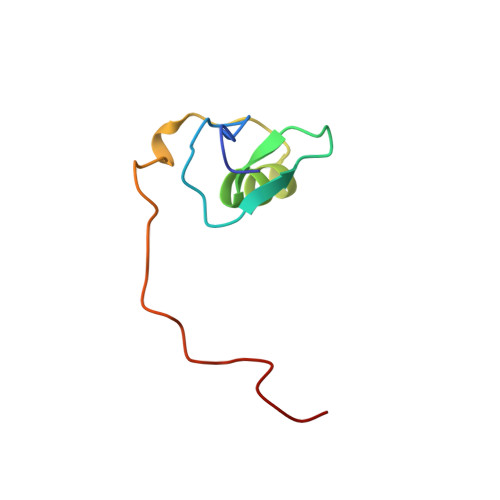
    2GAT, 3GAT

  • PubMed Abstract: 

    Anisotropy of the molecular magnetic susceptibility gives rise to a small degree of alignment. The resulting residual dipolar couplings, which can now be measured with the advent of higher magnetic fields in NMR, contain information on the orientation of the internuclear vectors relative to the molecular magnetic susceptibility tensor, thereby providing information on long range order that is not accessible by any of the solution NMR parameters currently used in structure determination. Thus, the dipolar couplings constitute unique and powerful restraints in determining the structures of magnetically oriented macromolecules in solution. The method is demonstrated on a complex of the DNA-binding domain of the transcription factor GATA-1 with a 16 base pair oligodeoxyribonucleotide.


  • Organizational Affiliation
    • Laboratory of Chemical Physics, National Institute of Diabetes and Digestive and Kidney Diseases, National Institutes of Health, Bethesda, Maryland 20892-0520, USA.

Macromolecule Content 

  • Total Structure Weight: 17.42 kDa 
  • Atom Count: 1,175 
  • Modeled Residue Count: 98 
  • Deposited Residue Count: 98 
  • Unique protein chains: 1
  • Unique nucleic acid chains: 2

Macromolecules


Find similar proteins by:|  3D Structure
Entity ID: 3
MoleculeChains  Sequence LengthOrganismDetailsImage
ERYTHROID TRANSCRIPTION FACTOR GATA-1C [auth A]66Gallus gallusMutation(s): 0 
UniProt
Find proteins for P17678 (Gallus gallus)
Explore P17678 
Go to UniProtKB:  P17678
Entity Groups
Sequence Clusters30% Identity50% Identity70% Identity90% Identity95% Identity100% Identity
UniProt GroupP17678
Sequence Annotations
Expand
Reference Sequence
Find similar nucleic acids by:  Sequence
Entity ID: 1
MoleculeChains LengthOrganismImage
DNA (5'-D(*GP*TP*TP*GP*CP*AP*GP*AP*TP*AP*AP*AP*CP*AP*TP*T)-3')A [auth B]16N/A
Sequence Annotations
Expand
Reference Sequence
Find similar nucleic acids by:  Sequence
Entity ID: 2
MoleculeChains LengthOrganismImage
DNA (5'-D(*AP*AP*TP*GP*TP*TP*TP*AP*TP*CP*TP*GP*CP*AP*AP*C)-3')B [auth C]16N/A
Sequence Annotations
Expand
Reference Sequence

Small Molecules

Ligands 1 Unique
IDChains Name / Formula / InChI Key2D Diagram3D Interactions
ZN

Query on ZN


Download:Ideal Coordinates CCD File
D [auth A]ZINC ION
Zn
PTFCDOFLOPIGGS-UHFFFAOYSA-N

Experimental Data & Validation

Experimental Data

  • Method: SOLUTION NMR
  • Conformers Calculated: 34 
  • Conformers Submitted: 
  • Selection Criteria: REGULARIZED MEAN STRUCTURE 

Structure Validation

View Full Validation Report


Entry History 

Deposition Data

Revision History  (Full details and data files)

  • Version 1.0: 1998-01-28
    Type: Initial release
  • Version 1.1: 2008-03-24
    Changes: Version format compliance
  • Version 1.2: 2011-07-13
    Changes: Version format compliance
  • Version 1.3: 2022-03-09
    Changes: Database references, Derived calculations, Other
  • Version 1.4: 2024-05-29
    Changes: Data collection