2GA8

Crystal structure of YFH7 from Saccharomyces cerevisiae: a putative P-loop containing kinase with a circular permutation.


Experimental Data Snapshot

  • Method: X-RAY DIFFRACTION
  • Resolution: 1.77 Å
  • R-Value Free: 0.219 
  • R-Value Work: 0.194 

wwPDB Validation   3D Report Full Report


This is version 1.2 of the entry. See complete history


Literature

Crystal structure and functional analysis identify the P-loop containing protein YFH7 of Saccharomyces cerevisiae as an ATP-dependent kinase.

Gueguen-Chaignon, V.Chaptal, V.Lariviere, L.Costa, N.Lopes, P.Morera, S.Nessler, S.

(2007) Proteins 71: 804-812

  • DOI: https://doi.org/10.1002/prot.21740
  • Primary Citation of Related Structures:  
    2GA8, 2GAA

  • PubMed Abstract: 

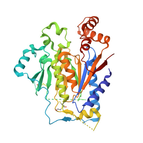
    Genome sequencing projects have revealed that P-loop proteins are highly represented in all organisms and that many of them have no attributed function. They are characterized by a conserved nucleotide-binding domain and carry different activities implicated in many cellular processes. Saccharomyces cerevisiae YFH7 is one of these P-loop proteins of unknown function. In this work we tried to integrate bioinformatics, structure, and enzymology to discover the function of YFH7. Sequence analysis revealed that yeast YFH7 is a yeast-specific protein showing weak similarity with the phosphoribulokinase/uridine kinase/bacterial pantothenate kinase (PRK/URK/PANK) subfamily of P-loop containing kinases. A large insertion of about 100 residues distinguishes YFH7 from other members of the family. The 1.95 A resolution crystal structure of YFH7 solved using the SAD method confirmed that YFH7 has a fold similar to the PRK/URK/PANK family, with the characteristic core, lid, and NMP(bind) domains. An additional alpha/beta domain of novel topology corresponds to the large sequence insertion. Structural and ligand binding analysis combined with enzymatic assays suggest that YFH7 is an ATP-dependent small molecule kinase with new substrate specificity.


  • Organizational Affiliation

    Laboratoire d'Enzymologie et Biochimie Structurales, CNRS, 1 av. de la terrasse, 91198 Gif-sur Yvette, France.


Macromolecules
Find similar proteins by:  (by identity cutoff)  |  3D Structure
Entity ID: 1
MoleculeChains Sequence LengthOrganismDetailsImage
Hypothetical 39.9 kDa protein359Saccharomyces cerevisiaeMutation(s): 3 
UniProt
Find proteins for P43591 (Saccharomyces cerevisiae (strain ATCC 204508 / S288c))
Explore P43591 
Go to UniProtKB:  P43591
Entity Groups  
Sequence Clusters30% Identity50% Identity70% Identity90% Identity95% Identity100% Identity
UniProt GroupP43591
Sequence Annotations
Expand
  • Reference Sequence
Small Molecules
Ligands 3 Unique
IDChains Name / Formula / InChI Key2D Diagram3D Interactions
HG
Query on HG

Download Ideal Coordinates CCD File 
B [auth A],
C [auth A],
D [auth A],
J [auth A]
MERCURY (II) ION
Hg
BQPIGGFYSBELGY-UHFFFAOYSA-N
SO4
Query on SO4

Download Ideal Coordinates CCD File 
E [auth A],
F [auth A],
G [auth A],
H [auth A],
I [auth A]
SULFATE ION
O4 S
QAOWNCQODCNURD-UHFFFAOYSA-L
BME
Query on BME

Download Ideal Coordinates CCD File 
K [auth A]BETA-MERCAPTOETHANOL
C2 H6 O S
DGVVWUTYPXICAM-UHFFFAOYSA-N
Modified Residues  1 Unique
IDChains TypeFormula2D DiagramParent
CME
Query on CME
A
L-PEPTIDE LINKINGC5 H11 N O3 S2CYS
Experimental Data & Validation

Experimental Data

  • Method: X-RAY DIFFRACTION
  • Resolution: 1.77 Å
  • R-Value Free: 0.219 
  • R-Value Work: 0.194 
  • Space Group: H 3
Unit Cell:
Length ( Å )Angle ( ˚ )
a = 135.278α = 90
b = 135.278β = 90
c = 48.476γ = 120
Software Package:
Software NamePurpose
DENZOdata reduction
SCALEPACKdata scaling
SOLVEphasing
CNSrefinement

Structure Validation

View Full Validation Report


Entry History 

Deposition Data

Revision History  (Full details and data files)

  • Version 1.0: 2007-03-20
    Type: Initial release
  • Version 1.1: 2007-12-12
    Changes: Version format compliance
  • Version 1.2: 2011-07-13
    Changes: Version format compliance