2GA4 | pdb_00002ga4

Stx2 with adenine


Experimental Data Snapshot

  • Method: X-RAY DIFFRACTION
  • Resolution: 1.80 Å
  • R-Value Free: 
    0.183 (Depositor), 0.180 (DCC) 
  • R-Value Work: 
    0.156 (Depositor), 0.153 (DCC) 
  • R-Value Observed: 
    0.157 (Depositor) 

wwPDB Validation 3D Report Full Report

Validation slider image for 2GA4

Ligand Structure Quality Assessment 


This is version 1.4 of the entry. See complete history

Literature

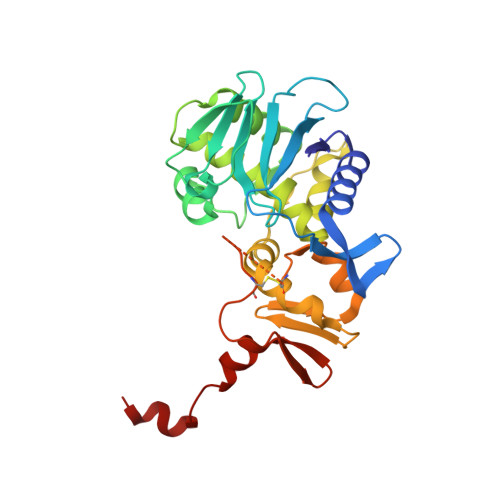
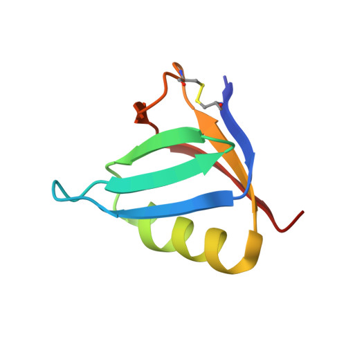
Binding of adenine to Stx2, the protein toxin from Escherichia coli O157:H7.

Fraser, M.E.Cherney, M.M.Marcato, P.Mulvey, G.L.Armstrong, G.D.James, M.N.

(2006) Acta Crystallogr Sect F Struct Biol Cryst Commun 62: 627-630

  • DOI: https://doi.org/10.1107/S1744309106021968
  • Primary Citation Related Structures: 
    2GA4

  • PubMed Abstract: 

    Stx2 is a protein toxin whose catalytic subunit acts as an N-glycosidase to depurinate a specific adenine base from 28S rRNA. In the holotoxin, the catalytic portion, A1, is linked to the rest of the A subunit, A2, and A2 interacts with the pentameric ring formed by the five B subunits. In order to test whether the holotoxin is active as an N-glycosidase, Stx2 was crystallized in the presence of adenosine and adenine. The crystals diffracted to approximately 1.8 angstroms and showed clear electron density for adenine in the active site. Adenosine had been cleaved, proving that Stx2 is an active N-glycosidase. While the holotoxin is active against small substrates, it would be expected that the B subunits would interfere with the binding of the 28S rRNA.


  • Organizational Affiliation
    • Department of Biological Sciences, University of Calgary, 2500 University Drive NW, Calgary AB T2N 1N4, Canada. frasm@ucalgary.ca

Macromolecule Content 

  • Total Structure Weight: 73.7 kDa 
  • Atom Count: 5,605 
  • Modeled Residue Count: 633 
  • Deposited Residue Count: 647 
  • Unique protein chains: 2

Macromolecules

Find similar proteins by:|  3D Structure
Entity ID: 1
MoleculeChains  Sequence LengthOrganismDetailsImage
Shiga-like toxin II subunit A297Escherichia phage 933WMutation(s): 0 
EC: 3.2.2.22
UniProt
Find proteins for P09385 (Escherichia phage 933W)
Explore P09385 
Go to UniProtKB:  P09385
Entity Groups
Sequence Clusters30% Identity50% Identity70% Identity90% Identity95% Identity100% Identity
UniProt GroupP09385
Sequence Annotations
Expand
Reference Sequence
Find similar proteins by:|  3D Structure
Entity ID: 2
MoleculeChains  Sequence LengthOrganismDetailsImage
Shiga-like toxin II subunit B
B, C, D, E, F
70Escherichia phage 933WMutation(s): 0 
UniProt
Find proteins for P09386 (Escherichia phage 933W)
Explore P09386 
Go to UniProtKB:  P09386
Entity Groups
Sequence Clusters30% Identity50% Identity70% Identity90% Identity95% Identity100% Identity
UniProt GroupP09386
Sequence Annotations
Expand
Reference Sequence

Small Molecules

Ligands 5 Unique
IDChains Name / Formula / InChI Key2D Diagram3D Interactions
1PS

Query on 1PS


Download:Ideal Coordinates CCD File
N [auth B],
O [auth C],
Q [auth D],
U [auth F]
3-PYRIDINIUM-1-YLPROPANE-1-SULFONATE
C8 H11 N O3 S
REEBJQTUIJTGAL-UHFFFAOYSA-N
ADE

Query on ADE


Download:Ideal Coordinates CCD File
J [auth A]ADENINE
C5 H5 N5
GFFGJBXGBJISGV-UHFFFAOYSA-N
EDO

Query on EDO


Download:Ideal Coordinates CCD File
V [auth F]1,2-ETHANEDIOL
C2 H6 O2
LYCAIKOWRPUZTN-UHFFFAOYSA-N
FMT

Query on FMT


Download:Ideal Coordinates CCD File
K [auth A],
L [auth A],
P [auth C],
R [auth D],
S [auth E]
FORMIC ACID
C H2 O2
BDAGIHXWWSANSR-UHFFFAOYSA-N
NA

Query on NA


Download:Ideal Coordinates CCD File
G [auth A],
H [auth A],
I [auth A],
M [auth B],
T [auth F]
SODIUM ION
Na
FKNQFGJONOIPTF-UHFFFAOYSA-N

Experimental Data & Validation

Experimental Data

  • Method: X-RAY DIFFRACTION
  • Resolution: 1.80 Å
  • R-Value Free:  0.183 (Depositor), 0.180 (DCC) 
  • R-Value Work:  0.156 (Depositor), 0.153 (DCC) 
  • R-Value Observed: 0.157 (Depositor) 
Space Group: P 61
Unit Cell:
Length ( Å )Angle ( ˚ )
a = 144.55α = 90
b = 144.55β = 90
c = 59.42γ = 120
Software Package:
Software NamePurpose
DENZOdata reduction
SCALEPACKdata scaling
CNSrefinement
PDB_EXTRACTdata extraction
XFITdata reduction

Structure Validation

View Full Validation Report


Ligand Structure Quality Assessment 


Entry History 

Deposition Data

  • Released Date: 2006-07-11 
  • Deposition Author(s): Fraser, M.E.

Revision History  (Full details and data files)

  • Version 1.0: 2006-07-11
    Type: Initial release
  • Version 1.1: 2008-05-01
    Changes: Version format compliance
  • Version 1.2: 2011-07-13
    Changes: Version format compliance
  • Version 1.3: 2017-10-18
    Changes: Refinement description
  • Version 1.4: 2024-10-30
    Changes: Data collection, Database references, Derived calculations, Structure summary