Enzyme Adaptation to Inhibitor Binding: A Cryptic Binding Site in Phenylethanolamine N-Methyltransferase
Gee, C.L., Drinkwater, N., Tyndall, J.D.A., Grunewald, G.L., Wu, Q., McLeish, M.J., Martin, J.L.(2007) J Med Chem 50: 4845-4853
- PubMed: 17845018 Search on PubMed
- DOI: https://doi.org/10.1021/jm0703385
- Primary Citation Related Structures:
2G70, 2G71, 2G72, 2OBF, 2ONY, 2ONZ, 2OPB - PubMed Abstract:
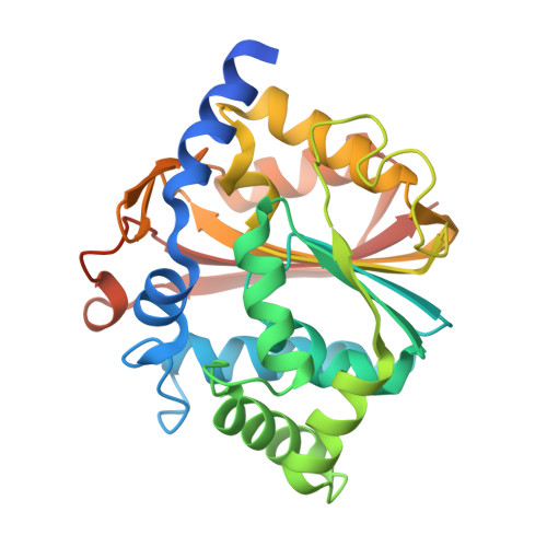
Shape complementarity is a fundamental principle of inhibitor design. Here we show that an enzyme for which the crystal structure has been determined (phenylethanolamine N-methyltransferase, PNMT) conceals a cryptic binding site. This site is revealed upon binding of inhibitors that are double the size of the physiological substrate. These large inhibitors are not predicted to bind in that they protrude through the accessible surface calculated from a PNMT/7-aminosulfonyl-1,2,3,4-tetrahydroisoquinoline (SK&F 29661) crystal structure, yet they are potent inhibitors of PNMT. We determined structures of the enzyme complexed with large inhibitors and found that the volume of the active site increases by 140 A3 upon binding. Changes in active site size and shape are brought about by unfavorable side chain conformations and rigid body helix motions. The energetic cost is modest, estimated at 2-3 kcal/mol from mutational analyses. Our findings further underline the importance of protein flexibility in structure-based inhibitor design studies.
- Institute for Molecular Bioscience and ARC Special Research Centre for Functional and Applied Genomics, University of Queensland, Brisbane, Qld, 4072 Australia.
Organizational Affiliation: