2G5B

Crystal Structure of the anti-Bax monoclonal antibody 6A7 and a Bax peptide.


Experimental Data Snapshot

  • Method: X-RAY DIFFRACTION
  • Resolution: 2.30 Å
  • R-Value Free: 0.275 
  • R-Value Work: 0.234 

wwPDB Validation   3D Report Full Report


This is version 2.0 of the entry. See complete history


Literature

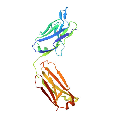
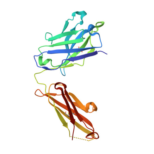
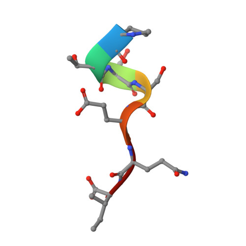
Elucidation of some Bax conformational changes through crystallization of an antibody-peptide complex.

Peyerl, F.W.Dai, S.Murphy, G.A.Crawford, F.White, J.Marrack, P.Kappler, J.W.

(2007) Cell Death Differ 14: 447-452

  • DOI: https://doi.org/10.1038/sj.cdd.4402025
  • Primary Citation of Related Structures:  
    2G5B

  • PubMed Abstract: 

    The Bcl-2 family member Bax plays a critical role in apoptosis. In healthy resting cells, Bax resides in the cytoplasm and loosely attached to the mitochondrial membrane. Apoptotic stimuli induce Bax activation, which is characterized by translocation and multimerization on the mitochondrial membrane surface resulting in exposure of an amino terminal epitope recognized by the monoclonal antibody 6A7. To understand the structural changes that occur during Bax activation, we determined the crystal structure of a Bax peptide bound to the 6A7 Fab fragment to a resolution of 2.3 A. The structure reveals the conformation of the 6A7 peptide epitope on Bax in the activated form and elucidates the extensive structural changes that Bax must undergo during the conversion from its native to its activated conformation.


  • Organizational Affiliation

    1Howard Hughes Medical Institute, Integrated Department of Immunology, Zuckerman Family/Canyon Ranch Crystallography Laboratory, National Jewish Medical and Research Center, Denver, CO 80206, USA.


Macromolecules
Find similar proteins by:  (by identity cutoff)  |  3D Structure
Entity ID: 1
MoleculeChains Sequence LengthOrganismDetailsImage
6A7 Fab Light Chain
A, C, E, G
217Mus musculusMutation(s): 0 
Entity Groups  
Sequence Clusters30% Identity50% Identity70% Identity90% Identity95% Identity100% Identity
Sequence Annotations
Expand
  • Reference Sequence
Find similar proteins by:  (by identity cutoff)  |  3D Structure
Entity ID: 2
MoleculeChains Sequence LengthOrganismDetailsImage
6A7 Fab Heavy Chain
B, D, F, H
222Mus musculusMutation(s): 0 
UniProt
Find proteins for P84751 (Mus musculus)
Explore P84751 
Go to UniProtKB:  P84751
Entity Groups  
Sequence Clusters30% Identity50% Identity70% Identity90% Identity95% Identity100% Identity
UniProt GroupP84751
Sequence Annotations
Expand
  • Reference Sequence

Find similar proteins by:  Sequence   |   3D Structure  

Entity ID: 3
MoleculeChains Sequence LengthOrganismDetailsImage
Bax Peptide
I, J, K, L
7N/AMutation(s): 0 
UniProt & NIH Common Fund Data Resources
Find proteins for Q07812 (Homo sapiens)
Explore Q07812 
Go to UniProtKB:  Q07812
PHAROS:  Q07812
GTEx:  ENSG00000087088 
Entity Groups  
UniProt GroupQ07812
Sequence Annotations
Expand
  • Reference Sequence
Oligosaccharides

Help

Entity ID: 4
MoleculeChains Length2D Diagram Glycosylation3D Interactions
beta-D-mannopyranose-(1-4)-2-acetamido-2-deoxy-beta-D-glucopyranose-(1-4)-2-acetamido-2-deoxy-beta-D-glucopyranose
M, O
3N-Glycosylation
Glycosylation Resources
GlyTouCan:  G15407YE
GlyCosmos:  G15407YE
GlyGen:  G15407YE
Entity ID: 5
MoleculeChains Length2D Diagram Glycosylation3D Interactions
2-acetamido-2-deoxy-alpha-D-glucopyranose-(1-4)-2-acetamido-2-deoxy-beta-D-glucopyranose
N
2N-Glycosylation
Glycosylation Resources
GlyTouCan:  G07375KG
GlyCosmos:  G07375KG
GlyGen:  G07375KG
Entity ID: 6
MoleculeChains Length2D Diagram Glycosylation3D Interactions
alpha-D-mannopyranose-(1-4)-2-acetamido-2-deoxy-beta-D-glucopyranose-(1-4)-2-acetamido-2-deoxy-beta-D-glucopyranose
P
3N-Glycosylation
Glycosylation Resources
GlyTouCan:  G62182OO
GlyCosmos:  G62182OO
GlyGen:  G62182OO
Experimental Data & Validation

Experimental Data

  • Method: X-RAY DIFFRACTION
  • Resolution: 2.30 Å
  • R-Value Free: 0.275 
  • R-Value Work: 0.234 
  • Space Group: P 21 21 2
Unit Cell:
Length ( Å )Angle ( ˚ )
a = 166.528α = 90
b = 183.897β = 90
c = 67.081γ = 90
Software Package:
Software NamePurpose
CNSrefinement
PDB_EXTRACTdata extraction
DENZOdata reduction
SCALEPACKdata scaling
AMoREphasing

Structure Validation

View Full Validation Report


Entry History 

Deposition Data

Revision History  (Full details and data files)

  • Version 1.0: 2006-07-25
    Type: Initial release
  • Version 1.1: 2008-05-01
    Changes: Version format compliance
  • Version 1.2: 2011-07-13
    Changes: Non-polymer description, Version format compliance
  • Version 2.0: 2020-07-29
    Type: Remediation
    Reason: Carbohydrate remediation
    Changes: Atomic model, Data collection, Derived calculations, Structure summary