2G3W | pdb_00002g3w

The Crystal Structure of YaeQ Protein from Xanthomonas axonopodis pv. citri


Experimental Data Snapshot

  • Method: X-RAY DIFFRACTION
  • Resolution: 1.90 Å
  • R-Value Free: 
    0.247 (Depositor), 0.247 (DCC) 
  • R-Value Work: 
    0.192 (Depositor), 0.192 (DCC) 
  • R-Value Observed: 
    0.194 (Depositor) 

wwPDB Validation 3D Report Full Report

Validation slider image for 2G3W

This is version 1.3 of the entry. See complete history

Literature

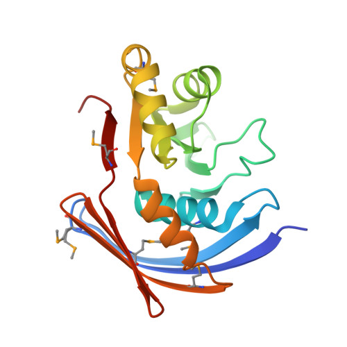
Structure of Xanthomonas axonopodis pv. citri YaeQ reveals a new compact protein fold built around a variation of the PD-(D/E)XK nuclease motif

Guzzo, C.R.Nagem, R.A.P.Barbosa, J.A.R.G.Farah, C.S.

(2007) Proteins 69: 644-651

  • DOI: https://doi.org/10.1002/prot.21556
  • Primary Citation Related Structures: 
    2G3W

  • PubMed Abstract: 

    The YaeQ family of proteins are found in many Gram-negative and a few Gram-positive bacteria. We have determined the first structure of a member of the YaeQ family by X-ray crystallography. Comparisons with other structures indicate that YaeQ represents a new compact protein fold built around a variation of the PD-(D/E)XK nuclease motif found in type II endonucleases and enzymes involved in DNA replication, repair, and recombination. We show that catalytically important residues in the PD-(D/E)XK nuclease superfamily are spatially conserved in YaeQ and other highly conserved YaeQ residues may be poised to interact with nucleic acid structures.


  • Organizational Affiliation
    • Departamento de Bioquímica, Instituto de Química, Universidade de São Paulo, Avenida Prof. Lineu Prestes 748, São Paulo, SP, CEP 05508-000, Brazil.

Macromolecule Content 

  • Total Structure Weight: 42.1 kDa 
  • Atom Count: 3,091 
  • Modeled Residue Count: 356 
  • Deposited Residue Count: 364 
  • Unique protein chains: 1

Macromolecules

Find similar proteins by:|  3D Structure
Entity ID: 1
MoleculeChains  Sequence LengthOrganismDetailsImage
hypothetical protein XAC2396
A, B
182Xanthomonas citriMutation(s): 0 
Gene Names: XAC2396

Small Molecules

Modified Residues  1 Unique
IDChains TypeFormula2D DiagramParent
MSE
Query on MSE
A, B
L-PEPTIDE LINKINGC5 H11 N O2 SeMET

Experimental Data & Validation

Experimental Data

  • Method: X-RAY DIFFRACTION
  • Resolution: 1.90 Å
  • R-Value Free:  0.247 (Depositor), 0.247 (DCC) 
  • R-Value Work:  0.192 (Depositor), 0.192 (DCC) 
  • R-Value Observed: 0.194 (Depositor) 
Space Group: P 1 21 1
Unit Cell:
Length ( Å )Angle ( ˚ )
a = 39.766α = 90
b = 91.969β = 108.35
c = 48.031γ = 90
Software Package:
Software NamePurpose
REFMACrefinement
HKL-2000data reduction
SCALEPACKdata scaling
SHARPphasing

Structure Validation

View Full Validation Report


Entry History 

Deposition Data

Revision History  (Full details and data files)

  • Version 1.0: 2007-02-27
    Type: Initial release
  • Version 1.1: 2008-05-01
    Changes: Version format compliance
  • Version 1.2: 2011-07-13
    Changes: Advisory, Version format compliance
  • Version 1.3: 2024-11-06
    Changes: Data collection, Database references, Derived calculations, Structure summary