2G31 | pdb_00002g31

Human Nogo-A functional domain: nogo60


Experimental Data Snapshot

  • Method: SOLUTION NMR
  • Conformers Calculated: 200 
  • Conformers Submitted: 20 
  • Selection Criteria: target function 

wwPDB Validation 3D Report Full Report

Validation slider image for 2G31

This is version 1.4 of the entry. See complete history

Literature

Nogo goes in the pure water: solution structure of Nogo-60 and design of the structured and buffer-soluble Nogo-54 for enhancing CNS regeneration

Li, M.F.Liu, J.X.Song, J.X.

(2006) Protein Sci 15: 1835-1841

  • DOI: https://doi.org/10.1110/ps.062306906
  • Primary Citation Related Structures: 
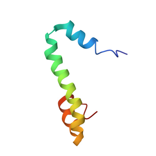
    2G31

  • PubMed Abstract: 

    The inability to determine the structure of the buffer-insoluble Nogo extracellular domain retarded further design of Nogo receptor (NgR) antagonists to treat CNS axonal injuries. Very surprisingly, we recently discovered that Nogo-60 was soluble and structured in salt-free water, thus allowing the determination of the first Nogo structure by heteronuclear NMR spectroscopy. Nogo-60 adopts an unusual helical structure with the N- and C-terminal helices connected by a long middle helix. While the N-helix has no contact with the rest of the molecule, the C-helix flips back to pack against the 20-residue middle helix. This packing appears to trigger the formation of the stable Nogo-60 structure because Nogo-40 with the last helix truncated is unstructured. The Nogo-60 structure offered us rationales for further design of the structured and buffer-soluble Nogo-54, which may be used as a novel NgR antagonist. Furthermore, our discovery may imply a general solution to solubilizing a category of buffer-insoluble proteins for urgent structural investigations.


  • Organizational Affiliation
    • Department of Biological Sciences, Faculty of Science, National University of Singapore, Singapore.

Macromolecule Content 

  • Total Structure Weight: 6.89 kDa 
  • Atom Count: 484 
  • Modeled Residue Count: 60 
  • Deposited Residue Count: 60 
  • Unique protein chains: 1

Macromolecules

Find similar proteins by:|  3D Structure
Entity ID: 1
MoleculeChains  Sequence LengthOrganismDetailsImage
Reticulon-460Homo sapiensMutation(s): 0 
Gene Names: NOGO
UniProt & NIH Common Fund Data Resources
Find proteins for Q9NQC3 (Homo sapiens)
Explore Q9NQC3 
Go to UniProtKB:  Q9NQC3
PHAROS:  Q9NQC3
GTEx:  ENSG00000115310 
Entity Groups
Sequence Clusters30% Identity50% Identity70% Identity90% Identity95% Identity100% Identity
UniProt GroupQ9NQC3
Sequence Annotations
Expand
Reference Sequence

Experimental Data & Validation

Experimental Data

  • Method: SOLUTION NMR
  • Conformers Calculated: 200 
  • Conformers Submitted: 20 
  • Selection Criteria: target function 

Structure Validation

View Full Validation Report


Entry History 

Deposition Data

Revision History  (Full details and data files)

  • Version 1.0: 2006-08-22
    Type: Initial release
  • Version 1.1: 2008-05-01
    Changes: Version format compliance
  • Version 1.2: 2011-07-13
    Changes: Version format compliance
  • Version 1.3: 2022-03-09
    Changes: Database references, Derived calculations
  • Version 1.4: 2024-05-29
    Changes: Data collection