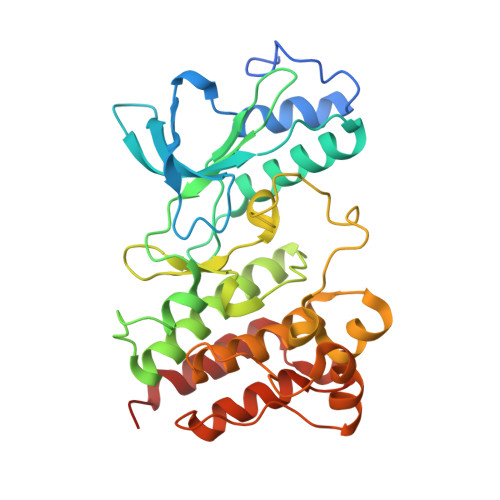
Structural characterization of autoinhibited c-Met kinase produced by coexpression in bacteria with phosphatase.
Wang, W., Marimuthu, A., Tsai, J., Kumar, A., Krupka, H.I., Zhang, C., Powell, B., Suzuki, Y., Nguyen, H., Tabrizizad, M., Luu, C., West, B.L.(2006) Proc Natl Acad Sci U S A 103: 3563-3568
- PubMed: 16537444 Search on PubMedSearch on PubMed Central
- DOI: https://doi.org/10.1073/pnas.0600048103
- Primary Citation Related Structures:
2G15 - PubMed Abstract:
Protein kinases are a large family of cell signaling mediators undergoing intensive research to identify inhibitors or modulators useful for medicine. As one strategy, small-molecule compounds that bind the active site with high affinity can be used to inhibit the enzyme activity. X-ray crystallography is a powerful method to reveal the structures of the kinase active sites, and thus aid in the design of high-affinity, selective inhibitors. However, a limitation still exists in the ability to produce purified kinases in amounts sufficient for crystallography. Furthermore, kinases exist in different conformation states as part of their normal regulation, and the ability to prepare crystals of kinases in these various states also remains a limitation. In this study, the c-Abl, c-Src, and c-Met kinases are produced in high yields in Escherichia coli by using a bicistronic vector encoding the PTP1B tyrosine phosphatase. A 100-fold lower dose of the inhibitor, Imatinib, was observed to inhibit the unphosphorylated form of c-Abl kinase prepared by using this vector, compared to the phosphorylated form produced without PTP1B, consistent with the known selectivity of this inhibitor for the unactivated conformation of the enzyme. Unphosphorylated c-Met kinase produced with this vector was used to obtain the crystal structure, at 2.15-A resolution, of the autoinhibited form of the kinase domain, revealing an intricate network of interactions involving c-Met residues documented previously to cause dysregulation when mutated in several cancers.
- Plexxikon, Inc., 91 Bolivar Drive, Berkeley, CA 94710, USA.
Organizational Affiliation: