2FXQ | pdb_00002fxq

Single-stranded DNA-binding protein from Thermus aquaticus


Experimental Data Snapshot

  • Method: X-RAY DIFFRACTION
  • Resolution: 1.85 Å
  • R-Value Free: 
    0.247 (Depositor), 0.246 (DCC) 
  • R-Value Work: 
    0.189 (Depositor), 0.189 (DCC) 
  • R-Value Observed: 
    0.192 (Depositor) 

wwPDB Validation 3D Report Full Report

Validation slider image for 2FXQ

This is version 1.3 of the entry. See complete history

Literature

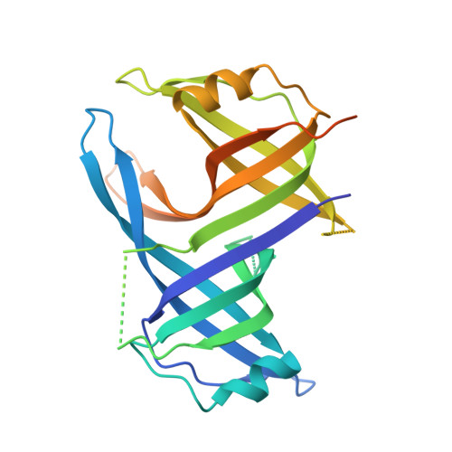
Structure of the single-stranded DNA-binding protein SSB from Thermus aquaticus.

Jedrzejczak, R.Dauter, M.Dauter, Z.Olszewski, M.Kur, J.

(2006) Acta Crystallogr D Biol Crystallogr 62: 1407-1412

  • DOI: https://doi.org/10.1107/S0907444906036031
  • Primary Citation Related Structures: 
    2FXQ

  • PubMed Abstract: 

    The crystal structure of the single-stranded DNA-binding protein from Thermus aquaticus has been solved and refined at 1.85 A resolution. Two monomers, each encompassing two oligonucleotide/oligosaccharide-binding (OB) domains and a number of flexible beta-hairpin loops, form an oligomer of approximate D(2) symmetry typical of bacterial SSBs. Comparison with other SSB structures confirms considerable variability in the mode of oligomerization and aggregation of SSB oligomers.


  • Organizational Affiliation
    • Synchrotron Radiation Research Section, MCL, National Cancer Institute, Argonne National Laboratory, Argonne, IL 60439, USA.

Macromolecule Content 

  • Total Structure Weight: 30.07 kDa 
  • Atom Count: 1,682 
  • Modeled Residue Count: 197 
  • Deposited Residue Count: 264 
  • Unique protein chains: 1

Macromolecules

Find similar proteins by:|  3D Structure
Entity ID: 1
MoleculeChains  Sequence LengthOrganismDetailsImage
Single-strand binding protein264Thermus aquaticusMutation(s): 0 
Gene Names: ssb
UniProt
Find proteins for Q9KH06 (Thermus aquaticus)
Explore Q9KH06 
Go to UniProtKB:  Q9KH06
Entity Groups
Sequence Clusters30% Identity50% Identity70% Identity90% Identity95% Identity100% Identity
UniProt GroupQ9KH06
Sequence Annotations
Expand
Reference Sequence

Experimental Data & Validation

Experimental Data

  • Method: X-RAY DIFFRACTION
  • Resolution: 1.85 Å
  • R-Value Free:  0.247 (Depositor), 0.246 (DCC) 
  • R-Value Work:  0.189 (Depositor), 0.189 (DCC) 
  • R-Value Observed: 0.192 (Depositor) 
Space Group: C 2 2 21
Unit Cell:
Length ( Å )Angle ( ˚ )
a = 51.14α = 90
b = 163.77β = 90
c = 60.16γ = 90
Software Package:
Software NamePurpose
REFMACrefinement
HKL-2000data reduction
SCALEPACKdata scaling
AMoREphasing

Structure Validation

View Full Validation Report


Entry History 

Deposition Data

Revision History  (Full details and data files)

  • Version 1.0: 2006-02-14
    Type: Initial release
  • Version 1.1: 2008-05-01
    Changes: Version format compliance
  • Version 1.2: 2011-07-13
    Changes: Advisory, Version format compliance
  • Version 1.3: 2024-02-14
    Changes: Data collection, Database references