High-resolution structures of Escherichia coli cDsbD in different redox states: A combined crystallographic, biochemical and computational study
Stirnimann, C.U., Rozhkova, A., Grauschopf, U., Boeckmann, R.A., Glockshuber, R., Capitani, G., Gruetter, M.G.(2006) J Mol Biology 358: 829-845
- PubMed: 16545842 Search on PubMed
- DOI: https://doi.org/10.1016/j.jmb.2006.02.030
- Primary Citation Related Structures:
2FWE, 2FWF, 2FWG, 2FWH - PubMed Abstract:
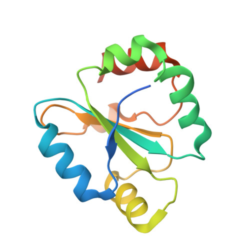
Escherichia coli DsbD transports electrons from cytoplasmic thioredoxin to periplasmic target proteins. DsbD is composed of an N-terminal (nDsbD) and a C-terminal (cDsbD) periplasmic domain, connected by a central transmembrane domain. Each domain possesses two cysteine residues essential for electron transport. The transport proceeds via disulfide exchange reactions from cytoplasmic thioredoxin to the central transmembrane domain and via cDsbD to nDsbD, which then reduces the periplasmic target proteins. We determined four high-resolution structures of cDsbD: oxidized (1.65 A resolution), chemically reduced (1.3 A), photo-reduced (1.1 A) and chemically reduced at pH increased from 4.6 to 7. The latter structure was refined at 0.99 A resolution, the highest achieved so far for a thioredoxin superfamily member. The data reveal unprecedented structural details of cDsbD, demonstrating that the domain is very rigid and undergoes hardly any conformational change upon disulfide reduction or interaction with nDsbD. In full agreement with the crystallographic results, guanidinium chloride-induced unfolding and refolding experiments indicate that oxidized and reduced cDsbD are equally stable. We confirmed the structural rigidity of cDsbD by molecular dynamics simulations. A remarkable feature of cDsbD is the pKa of 9.3 for the active site Cys461: this value, determined using two different experimental methods, surprisingly was around 2.5 units higher than expected on the basis of the redox potential. Additionally, taking advantage of the very high quality of the cDsbD structures, we carried out pKa calculations, which gave results in agreement with the experimental findings. In conclusion, our wide-scope analysis of cDsbD, encompassing atomic-resolution crystallography, computational chemistry and biophysical measurements, highlighted two so far unrecognized key aspects of this domain: its unusual redox properties and extreme rigidity. Both are likely to be correlated to the role of cDsbD as a covalently linked electron shuttle between the membrane domain and the N-terminal periplasmic domain of DsbD.
- Biochemisches Institut, Universität Zürich, Winterthurerstrasse 190, 8057 Zürich, Switzerland.
Organizational Affiliation: