2FUB | pdb_00002fub

Crystal structure of urate oxidase at 140 MPa


Experimental Data Snapshot

  • Method: X-RAY DIFFRACTION
  • Resolution: 2.30 Å
  • R-Value Free: 
    0.213 (Depositor), 0.158 (DCC) 
  • R-Value Work: 
    0.161 (Depositor), 0.165 (DCC) 
  • R-Value Observed: 
    0.164 (Depositor) 

Starting Model: experimental
View more details

wwPDB Validation 3D Report Full Report

Validation slider image for 2FUB

Ligand Structure Quality Assessment 


This is version 1.6 of the entry. See complete history

Literature

High pressure macromolecular crystallography: The 140-MPa crystal structure at 2.3 A resolution of urate oxidase, a 135-kDa tetrameric assembly

Colloc'h, N.Girard, E.Dhaussy, A.-C.Kahn, R.Ascone, I.Mezouar, M.Fourme, R.

(2006) Biochim Biophys Acta 1764: 391-397

  • DOI: https://doi.org/10.1016/j.bbapap.2006.01.006
  • Primary Citation Related Structures: 
    2FUB

  • PubMed Abstract: 

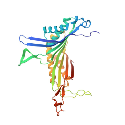
    We report the three-dimensional structure determined by high-pressure macromolecular crystallography (HPMX) of a 135-kDa homo-tetrameric enzyme, urate oxidase from Aspergillus flavus complexed with its potent inhibitor 8-azaxanthin. Urate oxidase crystals are quite sensitive to pressure, as three-dimensional order is lost at about 180 MPa. A highly complete 2.3 A resolution data set was collected at 140 MPa, close to the critical pressure. Crystal structures at atmospheric pressure and at high pressure were refined in the orthorhombic space group I222 with final crystallographic R factors 14.1% and 16.1%, respectively. The effect of pressure on temperature factors, ordered water molecules, hydrogen bond lengths, contacts, buried surface areas as well as cavity volume was investigated. Results suggest that the onset of disruption of the tetrameric assembly by pressure has been captured in the crystalline state.


  • Organizational Affiliation
    • Centre CYCERON, UMR 6185 Université de Caen-CNRS, Bd Becquerel, 14074 Caen, France.

Macromolecule Content 

  • Total Structure Weight: 34.43 kDa 
  • Atom Count: 2,500 
  • Modeled Residue Count: 295 
  • Deposited Residue Count: 301 
  • Unique protein chains: 1

Macromolecules

Find similar proteins by:|  3D Structure
Entity ID: 1
MoleculeChains  Sequence LengthOrganismDetailsImage
Uricase301Aspergillus flavusMutation(s): 0 
EC: 1.7.3.3
UniProt
Find proteins for Q00511 (Aspergillus flavus)
Explore Q00511 
Go to UniProtKB:  Q00511
Entity Groups
Sequence Clusters30% Identity50% Identity70% Identity90% Identity95% Identity100% Identity
UniProt GroupQ00511
Sequence Annotations
Expand
Reference Sequence

Experimental Data & Validation

Experimental Data

  • Method: X-RAY DIFFRACTION
  • Resolution: 2.30 Å
  • R-Value Free:  0.213 (Depositor), 0.158 (DCC) 
  • R-Value Work:  0.161 (Depositor), 0.165 (DCC) 
  • R-Value Observed: 0.164 (Depositor) 
Space Group: I 2 2 2
Unit Cell:
Length ( Å )Angle ( ˚ )
a = 79.18α = 90
b = 95.63β = 90
c = 104.65γ = 90
Software Package:
Software NamePurpose
REFMACrefinement
DENZOdata reduction
CCP4data scaling

Structure Validation

View Full Validation Report


Ligand Structure Quality Assessment 


Entry History 

Deposition Data

Revision History  (Full details and data files)

  • Version 1.0: 2006-02-14
    Type: Initial release
  • Version 1.1: 2008-05-01
    Changes: Version format compliance
  • Version 1.2: 2011-07-13
    Changes: Derived calculations, Version format compliance
  • Version 1.3: 2021-09-15
    Changes: Data collection, Database references, Derived calculations, Structure summary
  • Version 1.4: 2023-10-25
    Changes: Data collection, Refinement description
  • Version 1.5: 2023-11-15
    Changes: Derived calculations
  • Version 1.6: 2024-10-30
    Changes: Structure summary