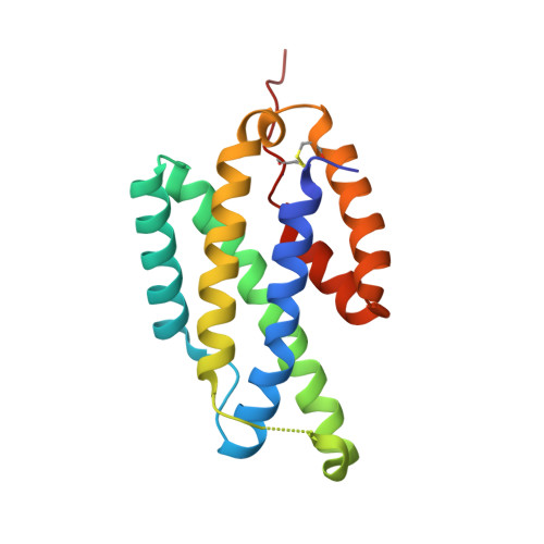
2FP2

Secreted Chorismate Mutase from Mycobacterium tuberculosis


Experimental Data Snapshot

  • Method: X-RAY DIFFRACTION
  • Resolution: 1.64 Å
  • R-Value Free: 0.203 
  • R-Value Work: 0.170 
  • R-Value Observed: 0.172 

Starting Model: experimental
View more details

wwPDB Validation   3D Report Full Report


Ligand Structure Quality Assessment 


This is version 1.4 of the entry. See complete history


Literature

1.6A Crystal Structure of the Secreted Chorismate Mutase from Mycobacterium tuberculosis: Novel Fold Topology Revealed

Okvist, M.Dey, R.Sasso, S.Grahn, E.Kast, P.Krengel, U.

(2006) J Mol Biol 357: 1483-1499

  • DOI: https://doi.org/10.1016/j.jmb.2006.01.069
  • Primary Citation of Related Structures:  
    2FP1, 2FP2

  • PubMed Abstract: 

    The presence of exported chorismate mutases produced by certain organisms such as Mycobacterium tuberculosis has been shown to correlate with their pathogenicity. As such, these proteins comprise a new group of promising selective drug targets. Here, we report the high-resolution crystal structure of the secreted dimeric chorismate mutase from M. tuberculosis (*MtCM; encoded by Rv1885c), which represents the first 3D-structure of a member of this chorismate mutase family, termed the AroQ(gamma) subclass. Structures are presented both for the unliganded enzyme and for a complex with a transition state analog. The protomer fold resembles the structurally characterized (dimeric) Escherichia coli chorismate mutase domain, but exhibits a new topology, with helix H4 of *MtCM carrying the catalytic site residue missing in the shortened helix H1. Furthermore, the structure of each *MtCM protomer is significantly more compact and only harbors one active site pocket, which is formed entirely by one polypeptide chain. Apart from the structural model, we present evidence as to how the substrate may enter the active site.


  • Organizational Affiliation

    Department of Chemistry and Bioscience, Chalmers University of Technology, P.O. Box 462, SE-40530 Göteborg, Sweden.


Macromolecules
Find similar proteins by:  (by identity cutoff)  |  3D Structure
Entity ID: 1
MoleculeChains Sequence LengthOrganismDetailsImage
Chorismate mutase
A, B
166Mycobacterium tuberculosisMutation(s): 0 
EC: 5.4.99.5
UniProt
Find proteins for P9WIB9 (Mycobacterium tuberculosis (strain ATCC 25618 / H37Rv))
Explore P9WIB9 
Go to UniProtKB:  P9WIB9
Entity Groups  
Sequence Clusters30% Identity50% Identity70% Identity90% Identity95% Identity100% Identity
UniProt GroupP9WIB9
Sequence Annotations
Expand
  • Reference Sequence
Small Molecules
Ligands 1 Unique
IDChains Name / Formula / InChI Key2D Diagram3D Interactions
TSA
Query on TSA

Download Ideal Coordinates CCD File 
C [auth B]8-HYDROXY-2-OXA-BICYCLO[3.3.1]NON-6-ENE-3,5-DICARBOXYLIC ACID
C10 H12 O6
KRZHNRULRHECRF-JQCUSGDOSA-N
Experimental Data & Validation

Experimental Data

  • Method: X-RAY DIFFRACTION
  • Resolution: 1.64 Å
  • R-Value Free: 0.203 
  • R-Value Work: 0.170 
  • R-Value Observed: 0.172 
  • Space Group: P 1 21 1
Unit Cell:
Length ( Å )Angle ( ˚ )
a = 42.906α = 90
b = 72.8β = 103.99
c = 61.716γ = 90
Software Package:
Software NamePurpose
REFMACrefinement
MOSFLMdata reduction
CCP4data scaling
MOLREPphasing

Structure Validation

View Full Validation Report


Ligand Structure Quality Assessment 


Entry History 

Deposition Data

Revision History  (Full details and data files)

  • Version 1.0: 2006-03-28
    Type: Initial release
  • Version 1.1: 2008-05-01
    Changes: Version format compliance
  • Version 1.2: 2011-07-13
    Changes: Version format compliance
  • Version 1.3: 2018-03-07
    Changes: Data collection
  • Version 1.4: 2023-08-30
    Changes: Data collection, Database references, Derived calculations, Refinement description