2FMD | pdb_00002fmd

Structural basis of carbohydrate recognition by Bowringia milbraedii seed agglutinin


Experimental Data Snapshot

  • Method: X-RAY DIFFRACTION
  • Resolution: 1.90 Å
  • R-Value Free: 
    0.228 (Depositor), 0.227 (DCC) 
  • R-Value Work: 
    0.183 (Depositor), 0.182 (DCC) 
  • R-Value Observed: 
    0.186 (Depositor) 

Starting Model: experimental
View more details

wwPDB Validation 3D Report Full Report

Validation slider image for 2FMD

This is version 2.2 of the entry. See complete history

Literature

Structural basis of carbohydrate recognition by a Man(alpha1-2)Man-specific lectin from Bowringia milbraedii.

Buts, L.Garcia-Pino, A.Wyns, L.Loris, R.

(2006) Glycobiology 16: 635-640

  • DOI: https://doi.org/10.1093/glycob/cwj109
  • Primary Citation Related Structures: 
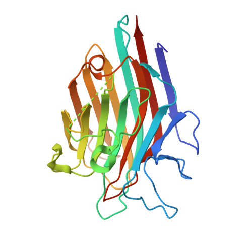
    2FMD

  • PubMed Abstract: 

    The crystal structure of the seed lectin from the tropical legume Bowringia milbraedii was determined in complex with the disaccharide ligand Man(alpha1-2)Man. In solution, the protein exhibits a dynamic dimer-tetramer equilibrium, consistent with the concanavalin A-type tetramer observed in the crystal. Contacts between the tetramers are mediated almost exclusively through the carbohydrate ligand, resulting in a crystal lattice virtually identical to that of the concanavalin-A:Man(alpha1-2)Man complex, even though both proteins have less than 50% sequence identity. The disaccharide binds exclusively in a "downstream" binding mode, with the non-reducing mannose occupying the monosaccharide-binding site. The reducing mannose is bound in a predominantly polar subsite involving Tyr131, Gln218, and Tyr219.


  • Organizational Affiliation
    • Laboratorium voor Ultrastructuur, Vrije Universiteit Brussel and Department of Molecular and Cellular Interactions, Vlaams Interuniversitair Instituut voor Biotechnologie, Pleinlaan 2, B-1050 Brussels, Belgium. lievbuts@vub.ac.be

Macromolecule Content 

  • Total Structure Weight: 26.02 kDa 
  • Atom Count: 1,974 
  • Modeled Residue Count: 232 
  • Deposited Residue Count: 240 
  • Unique protein chains: 1

Macromolecules

Find similar proteins by:|  3D Structure
Entity ID: 1
MoleculeChains  Sequence LengthOrganismDetailsImage
Lectin240Leucomphalos mildbraediiMutation(s): 0 
UniProt
Find proteins for P42088 (Leucomphalos mildbraedii)
Explore P42088 
Go to UniProtKB:  P42088
Entity Groups
Sequence Clusters30% Identity50% Identity70% Identity90% Identity95% Identity100% Identity
UniProt GroupP42088
Sequence Annotations
Expand
Reference Sequence

Oligosaccharides

Help  
Entity ID: 2
MoleculeChains Length2D Diagram GlycosylationD Interactions
alpha-D-mannopyranose-(1-2)-alpha-D-mannopyranose
B
2N/A
Glycosylation Resources
GlyTouCan: G53402KW
GlyCosmos: G53402KW
GlyGen: G53402KW

Biologically Interesting Molecules (External Reference) 

1 Unique

Experimental Data & Validation

Experimental Data

  • Method: X-RAY DIFFRACTION
  • Resolution: 1.90 Å
  • R-Value Free:  0.228 (Depositor), 0.227 (DCC) 
  • R-Value Work:  0.183 (Depositor), 0.182 (DCC) 
  • R-Value Observed: 0.186 (Depositor) 
Space Group: I 2 2 2
Unit Cell:
Length ( Å )Angle ( ˚ )
a = 66.603α = 90
b = 86.351β = 90
c = 91.76γ = 90
Software Package:
Software NamePurpose
DENZOdata reduction
SCALEPACKdata scaling
PHASERphasing
CNSrefinement

Structure Validation

View Full Validation Report


Entry History 

Deposition Data

Revision History  (Full details and data files)

  • Version 1.0: 2006-08-22
    Type: Initial release
  • Version 1.1: 2008-05-01
    Changes: Version format compliance
  • Version 1.2: 2011-07-13
    Changes: Version format compliance
  • Version 2.0: 2020-07-29
    Type: Remediation
    Reason: Carbohydrate remediation
    Changes: Atomic model, Data collection, Derived calculations, Structure summary
  • Version 2.1: 2023-08-30
    Changes: Data collection, Database references, Refinement description, Structure summary
  • Version 2.2: 2024-11-13
    Changes: Structure summary