2FCU | pdb_00002fcu

SyrB2 with alpha-ketoglutarate


Experimental Data Snapshot

  • Method: X-RAY DIFFRACTION
  • Resolution: 1.60 Å
  • R-Value Free: 
    0.212 (Depositor), 0.215 (DCC) 
  • R-Value Work: 
    0.188 (Depositor), 0.193 (DCC) 
  • R-Value Observed: 
    0.189 (Depositor) 

wwPDB Validation 3D Report Full Report

Validation slider image for 2FCU

This is version 1.3 of the entry. See complete history

Literature

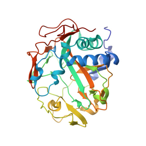
Crystal structure of the non-haem iron halogenase SyrB2 in syringomycin biosynthesis.

Blasiak, L.C.Vaillancourt, F.H.Walsh, C.T.Drennan, C.L.

(2006) Nature 440: 368-371

  • DOI: https://doi.org/10.1038/nature04544
  • Primary Citation Related Structures: 
    2FCT, 2FCU, 2FCV

  • PubMed Abstract: 

    Non-haem Fe(II)/alpha-ketoglutarate (alphaKG)-dependent enzymes harness the reducing power of alphaKG to catalyse oxidative reactions, usually the hydroxylation of unactivated carbons, and are involved in processes such as natural product biosynthesis, the mammalian hypoxic response, and DNA repair. These enzymes couple the decarboxylation of alphaKG with the formation of a high-energy ferryl-oxo intermediate that acts as a hydrogen-abstracting species. All previously structurally characterized mononuclear iron enzymes contain a 2-His, 1-carboxylate motif that coordinates the iron. The two histidines and one carboxylate, known as the 'facial triad', form one triangular side of an octahedral iron coordination geometry. A subclass of mononuclear iron enzymes has been shown to catalyse halogenation reactions, rather than the more typical hydroxylation reaction. SyrB2, a member of this subclass, is a non-haem Fe(II)/alphaKG-dependent halogenase that catalyses the chlorination of threonine in syringomycin E biosynthesis. Here we report the structure of SyrB2 with both a chloride ion and alphaKG coordinated to the iron ion at 1.6 A resolution. This structure reveals a previously unknown coordination of iron, in which the carboxylate ligand of the facial triad is replaced by a chloride ion.


  • Organizational Affiliation
    • Department of Chemistry, Massachusetts Institute of Technology, Cambridge, Massachusetts 02139, USA.

Macromolecule Content 

  • Total Structure Weight: 72.15 kDa 
  • Atom Count: 5,368 
  • Modeled Residue Count: 608 
  • Deposited Residue Count: 626 
  • Unique protein chains: 1

Macromolecules

Find similar proteins by:|  3D Structure
Entity ID: 1
MoleculeChains  Sequence LengthOrganismDetailsImage
syringomycin biosynthesis enzyme 2
A, B
313Pseudomonas syringae pv. syringaeMutation(s): 0 
Gene Names: SyrB2
EC: 1.14.20.15
UniProt
Find proteins for Q9RBY6 (Pseudomonas syringae pv. syringae)
Explore Q9RBY6 
Go to UniProtKB:  Q9RBY6
Entity Groups
Sequence Clusters30% Identity50% Identity70% Identity90% Identity95% Identity100% Identity
UniProt GroupQ9RBY6
Sequence Annotations
Expand
Reference Sequence

Small Molecules

Ligands 2 Unique
IDChains Name / Formula / InChI Key2D Diagram3D Interactions
DSU

Query on DSU


Download:Ideal Coordinates CCD File
C [auth A]((2R,3S,4S,5S)-3,4-DIHYDROXY-5-(HYDROXYMETHYL)-5-((2R,3S,4S,5S,6R)-3,4,5-TRIHYDROXY-6-METHOXY-TETRAHYDRO-2H-PYRAN-2-YLOXY)-TETRAHYDROFURAN-2-YL)METHYL NONANOATE
C22 H40 O12
GCKKEFVHVGTJPT-PZOSHJPSSA-N
AKG

Query on AKG


Download:Ideal Coordinates CCD File
D [auth A],
E [auth B]
2-OXOGLUTARIC ACID
C5 H6 O5
KPGXRSRHYNQIFN-UHFFFAOYSA-N

Experimental Data & Validation

Experimental Data

  • Method: X-RAY DIFFRACTION
  • Resolution: 1.60 Å
  • R-Value Free:  0.212 (Depositor), 0.215 (DCC) 
  • R-Value Work:  0.188 (Depositor), 0.193 (DCC) 
  • R-Value Observed: 0.189 (Depositor) 
Space Group: P 21 21 21
Unit Cell:
Length ( Å )Angle ( ˚ )
a = 57.263α = 90
b = 90.032β = 90
c = 128.769γ = 90
Software Package:
Software NamePurpose
SCALAdata scaling
REFMACrefinement
PDB_EXTRACTdata extraction
MOSFLMdata reduction
CCP4data scaling
SHARPphasing

Structure Validation

View Full Validation Report


Entry History 

Deposition Data

Revision History  (Full details and data files)

  • Version 1.0: 2006-03-21
    Type: Initial release
  • Version 1.1: 2008-05-01
    Changes: Version format compliance
  • Version 1.2: 2011-07-13
    Changes: Source and taxonomy, Version format compliance
  • Version 1.3: 2024-02-14
    Changes: Data collection, Database references, Derived calculations