2FCK | pdb_00002fck

Structure of a putative ribosomal-protein-serine acetyltransferase from Vibrio cholerae.


Experimental Data Snapshot

  • Method: X-RAY DIFFRACTION
  • Resolution: 1.70 Å
  • R-Value Free: 
    0.244 (Depositor), 0.241 (DCC) 
  • R-Value Work: 
    0.211 (Depositor), 0.210 (DCC) 
  • R-Value Observed: 
    0.212 (Depositor) 

wwPDB Validation 3D Report Full Report

Validation slider image for 2FCK

This is version 1.4 of the entry. See complete history

Literature

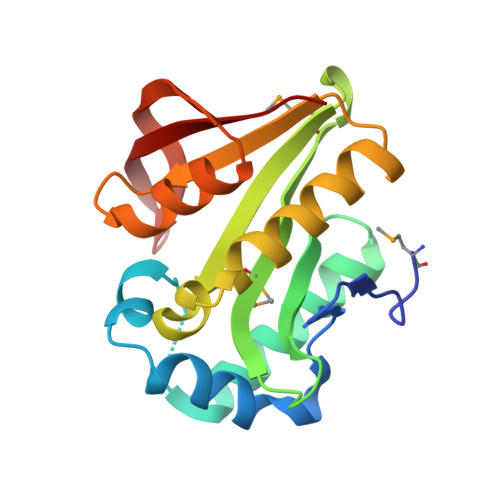
Crystal structure of an acetyltransferase protein from Vibrio cholerae strain N16961.

Cuff, M.E.Li, H.Moy, S.Watson, J.Cipriani, A.Joachimiak, A.

(2007) Proteins 69: 422-427

Macromolecule Content 

  • Total Structure Weight: 21.78 kDa 
  • Atom Count: 1,786 
  • Modeled Residue Count: 174 
  • Deposited Residue Count: 181 
  • Unique protein chains: 1

Macromolecules

Find similar proteins by:|  3D Structure
Entity ID: 1
MoleculeChains  Sequence LengthOrganismDetailsImage
ribosomal-protein-serine acetyltransferase, putative181Vibrio cholerae O1 biovar El Tor str. N16961Mutation(s): 3 
Gene Names: VC1889
UniProt
Find proteins for A0A0H3AIE8 (Vibrio cholerae serotype O1 (strain ATCC 39541 / Classical Ogawa 395 / O395))
Explore A0A0H3AIE8 
Go to UniProtKB:  A0A0H3AIE8
Entity Groups
Sequence Clusters30% Identity50% Identity70% Identity90% Identity95% Identity100% Identity
UniProt GroupA0A0H3AIE8
Sequence Annotations
Expand
Reference Sequence

Small Molecules

Ligands 2 Unique
IDChains Name / Formula / InChI Key2D Diagram3D Interactions
GOL

Query on GOL


Download:Ideal Coordinates CCD File
N [auth A]GLYCEROL
C3 H8 O3
PEDCQBHIVMGVHV-UHFFFAOYSA-N
NO3

Query on NO3


Download:Ideal Coordinates CCD File
B [auth A]
C [auth A]
D [auth A]
E [auth A]
F [auth A]
B [auth A],
C [auth A],
D [auth A],
E [auth A],
F [auth A],
G [auth A],
H [auth A],
I [auth A],
J [auth A],
K [auth A],
L [auth A],
M [auth A]
NITRATE ION
N O3
NHNBFGGVMKEFGY-UHFFFAOYSA-N
Modified Residues  1 Unique
IDChains TypeFormula2D DiagramParent
MSE
Query on MSE
A
L-PEPTIDE LINKINGC5 H11 N O2 SeMET

Experimental Data & Validation

Experimental Data

  • Method: X-RAY DIFFRACTION
  • Resolution: 1.70 Å
  • R-Value Free:  0.244 (Depositor), 0.241 (DCC) 
  • R-Value Work:  0.211 (Depositor), 0.210 (DCC) 
  • R-Value Observed: 0.212 (Depositor) 
Space Group: P 21 21 2
Unit Cell:
Length ( Å )Angle ( ˚ )
a = 53.051α = 90
b = 103.065β = 90
c = 37.861γ = 90
Software Package:
Software NamePurpose
REFMACrefinement
SBC-Collectdata collection
HKL-2000data scaling
PHENIXphasing
autoSHARPphasing
ARP/wARPmodel building

Structure Validation

View Full Validation Report


Entry History 

Deposition Data

Revision History  (Full details and data files)

  • Version 1.0: 2006-02-28
    Type: Initial release
  • Version 1.1: 2007-09-24
    Changes: Version format compliance
  • Version 1.2: 2011-07-13
    Changes: Advisory, Source and taxonomy, Version format compliance
  • Version 1.3: 2017-10-18
    Changes: Refinement description
  • Version 1.4: 2024-10-16
    Changes: Data collection, Database references, Derived calculations, Structure summary