2FBX | pdb_00002fbx

WRN exonuclease, Mg complex


Experimental Data Snapshot

  • Method: X-RAY DIFFRACTION
  • Resolution: 2.20 Å
  • R-Value Free: 
    0.262 (Depositor), 0.257 (DCC) 
  • R-Value Work: 
    0.242 (Depositor), 0.236 (DCC) 
  • R-Value Observed: 
    0.242 (Depositor) 

Starting Model: experimental
View more details

wwPDB Validation 3D Report Full Report

Validation slider image for 2FBX

This is version 1.4 of the entry. See complete history

Literature

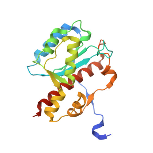
WRN exonuclease structure and molecular mechanism imply an editing role in DNA end processing.

Perry, J.J.Yannone, S.M.Holden, L.G.Hitomi, C.Asaithamby, A.Han, S.Cooper, P.K.Chen, D.J.Tainer, J.A.

(2006) Nat Struct Mol Biol 13: 414-422

  • DOI: https://doi.org/10.1038/nsmb1088
  • Primary Citation Related Structures: 
    2FBT, 2FBV, 2FBX, 2FBY, 2FC0

  • PubMed Abstract: 

    WRN is unique among the five human RecQ DNA helicases in having a functional exonuclease domain (WRN-exo) and being defective in the premature aging and cancer-related disorder Werner syndrome. Here, we characterize WRN-exo crystal structures, biochemical activity and participation in DNA end joining. Metal-ion complex structures, active site mutations and activity assays reveal a nuclease mechanism mediated by two metal ions. The DNA end-binding Ku70/80 complex specifically stimulates WRN-exo activity, and structure-based mutational inactivation of WRN-exo alters DNA end joining in human cells. We furthermore establish structural and biochemical similarities of WRN-exo to DnaQ-family replicative proofreading exonucleases, describing WRN-specific adaptations consistent with double-stranded DNA specificity and functionally important conformational changes. These results indicate WRN-exo is a human DnaQ family member and support DnaQ-like proofreading activities stimulated by Ku70/80, with implications for WRN functions in age-related pathologies and maintenance of genomic integrity.


  • Organizational Affiliation
    • Department of Molecular Biology and Skaggs Institute for Chemical Biology, The Scripps Research Institute, La Jolla, California 92037, USA.

Macromolecule Content 

  • Total Structure Weight: 23.48 kDa 
  • Atom Count: 1,760 
  • Modeled Residue Count: 195 
  • Deposited Residue Count: 205 
  • Unique protein chains: 1

Macromolecules

Find similar proteins by:|  3D Structure
Entity ID: 1
MoleculeChains  Sequence LengthOrganismDetailsImage
Werner syndrome helicase205Homo sapiensMutation(s): 0 
Gene Names: WRNRECQ3RECQL2
EC: 2.7.7 (PDB Primary Data), 5.6.2.4 (UniProt), 3.1 (UniProt)
UniProt & NIH Common Fund Data Resources
Find proteins for Q14191 (Homo sapiens)
Explore Q14191 
Go to UniProtKB:  Q14191
PHAROS:  Q14191
GTEx:  ENSG00000165392 
Entity Groups
Sequence Clusters30% Identity50% Identity70% Identity90% Identity95% Identity100% Identity
UniProt GroupQ14191
Sequence Annotations
Expand
Reference Sequence

Experimental Data & Validation

Experimental Data

  • Method: X-RAY DIFFRACTION
  • Resolution: 2.20 Å
  • R-Value Free:  0.262 (Depositor), 0.257 (DCC) 
  • R-Value Work:  0.242 (Depositor), 0.236 (DCC) 
  • R-Value Observed: 0.242 (Depositor) 
Space Group: P 32 2 1
Unit Cell:
Length ( Å )Angle ( ˚ )
a = 80.806α = 90
b = 80.806β = 90
c = 93.174γ = 120
Software Package:
Software NamePurpose
HKL-2000data collection
SCALEPACKdata scaling
AMoREphasing
CNSrefinement
HKL-2000data reduction

Structure Validation

View Full Validation Report


Entry History 

Deposition Data

  • Released Date: 2006-04-25 
  • Deposition Author(s): Perry, J.J.

Revision History  (Full details and data files)

  • Version 1.0: 2006-04-25
    Type: Initial release
  • Version 1.1: 2008-05-01
    Changes: Version format compliance
  • Version 1.2: 2011-07-13
    Changes: Version format compliance
  • Version 1.3: 2017-10-11
    Changes: Data collection
  • Version 1.4: 2023-08-30
    Changes: Data collection, Database references, Derived calculations, Refinement description