2F8J | pdb_00002f8j

Crystal structure of Histidinol-phosphate aminotransferase (EC 2.6.1.9) (Imidazole acetol-phosphate transferase) (tm1040) from Thermotoga maritima at 2.40 A resolution


Experimental Data Snapshot

  • Method: X-RAY DIFFRACTION
  • Resolution: 2.40 Å
  • R-Value Free: 
    0.238 (Depositor), 0.302 (DCC) 
  • R-Value Work: 
    0.182 (Depositor) 
  • R-Value Observed: 
    0.185 (Depositor) 

Starting Model: experimental
View more details

wwPDB Validation 3D Report Full Report

Validation slider image for 2F8J

Ligand Structure Quality Assessment 


This is version 1.4 of the entry. See complete history

Literature

Crystal structure of Histidinol-phosphate aminotransferase (EC 2.6.1.9) (Imidazole acetol-phosphate transferase) (tm1040) from Thermotoga maritima at 2.40 A resolution

Joint Center for Structural Genomics (JCSG)

To be published.

Macromolecule Content 

  • Total Structure Weight: 165.05 kDa 
  • Atom Count: 11,003 
  • Modeled Residue Count: 1,336 
  • Deposited Residue Count: 1,388 
  • Unique protein chains: 1

Macromolecules

Find similar proteins by:|  3D Structure
Entity ID: 1
MoleculeChains  Sequence LengthOrganismDetailsImage
Histidinol-phosphate aminotransferase
A, B, C, D
347Thermotoga maritimaMutation(s): 0 
Gene Names: hisC
EC: 2.6.1.9
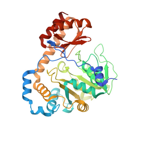
UniProt
Find proteins for Q9X0D0 (Thermotoga maritima (strain ATCC 43589 / DSM 3109 / JCM 10099 / NBRC 100826 / MSB8))
Explore Q9X0D0 
Go to UniProtKB:  Q9X0D0
Entity Groups
Sequence Clusters30% Identity50% Identity70% Identity90% Identity95% Identity100% Identity
UniProt GroupQ9X0D0
Sequence Annotations
Expand
Reference Sequence

Small Molecules

Ligands 2 Unique
IDChains Name / Formula / InChI Key2D Diagram3D Interactions
PMP

Query on PMP


Download:Ideal Coordinates CCD File
E [auth A],
I [auth B],
O [auth C],
S [auth D]
4'-DEOXY-4'-AMINOPYRIDOXAL-5'-PHOSPHATE
C8 H13 N2 O5 P
ZMJGSOSNSPKHNH-UHFFFAOYSA-N
EDO

Query on EDO


Download:Ideal Coordinates CCD File
F [auth A]
G [auth A]
H [auth A]
J [auth B]
K [auth B]
F [auth A],
G [auth A],
H [auth A],
J [auth B],
K [auth B],
L [auth B],
M [auth B],
N [auth B],
P [auth C],
Q [auth C],
R [auth C],
T [auth D],
U [auth D]
1,2-ETHANEDIOL
C2 H6 O2
LYCAIKOWRPUZTN-UHFFFAOYSA-N

Experimental Data & Validation

Experimental Data

  • Method: X-RAY DIFFRACTION
  • Resolution: 2.40 Å
  • R-Value Free:  0.238 (Depositor), 0.302 (DCC) 
  • R-Value Work:  0.182 (Depositor) 
  • R-Value Observed: 0.185 (Depositor) 
Space Group: P 21 21 2
Unit Cell:
Length ( Å )Angle ( ˚ )
a = 145.156α = 90
b = 187.453β = 90
c = 54.316γ = 90
Software Package:
Software NamePurpose
REFMACrefinement
SCALAdata scaling
PDB_EXTRACTdata extraction
DENZOdata reduction
SCALEPACKdata scaling
MOLREPphasing

Structure Validation

View Full Validation Report


Ligand Structure Quality Assessment 


Entry History 

Deposition Data

Revision History  (Full details and data files)

  • Version 1.0: 2006-02-21
    Type: Initial release
  • Version 1.1: 2008-05-01
    Changes: Version format compliance
  • Version 1.2: 2011-07-13
    Changes: Advisory, Version format compliance
  • Version 1.3: 2023-01-25
    Changes: Database references, Derived calculations
  • Version 1.4: 2023-09-20
    Changes: Data collection, Refinement description